Deposition Date
2002-06-05
Release Date
2002-10-30
Last Version Date
2024-02-14
Entry Detail
PDB ID:
1LXM
Keywords:
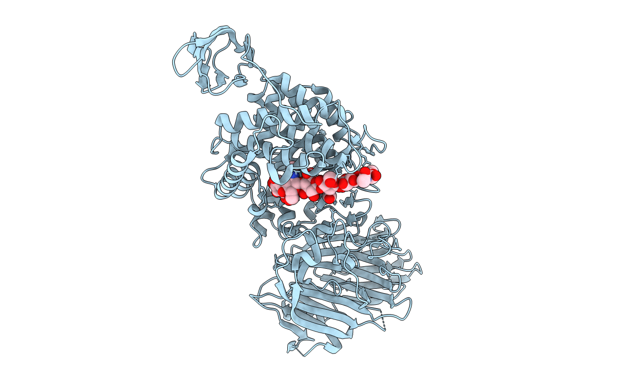
Title:
Crystal Structure of Streptococcus agalactiae Hyaluronate Lyase Complexed with Hexasaccharide Unit of Hyaluronan
Biological Source:
Source Organism(s):
Streptococcus agalactiae (Taxon ID: 1311)
Expression System(s):
Method Details:
Experimental Method:
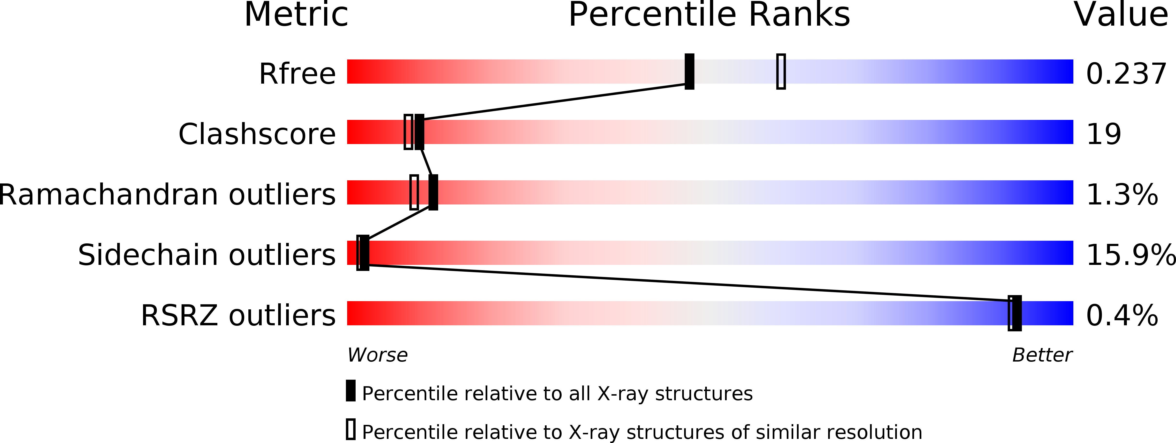
Resolution:
2.20 Å
R-Value Free:
0.27
R-Value Work:
0.21
R-Value Observed:
0.21
Space Group:
C 2 2 21