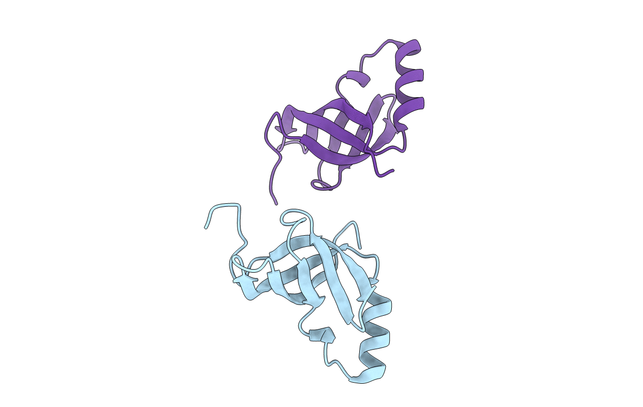
1LUZ image
Deposition Date 2002-05-23
Release Date 2002-08-28
Last Version Date 2024-10-30
Entry Detail
PDB ID:
1LUZ
Keywords:
Title:
Crystal Structure of the K3L Protein From Vaccinia Virus (Wisconsin Strain)
Biological Source:
Source Organism:
Vaccinia virus (Taxon ID: 10254)
Host Organism:
Method Details:
Experimental Method:
Resolution:
1.80 Å
R-Value Free:
0.25
R-Value Work:
0.23
Space Group:
C 1 2 1
Macromolecular Entities
Structures with similar UniProt ID
Protein Blast
Polymer Type:polypeptide(L)
Molecule:Protein K3
Gene (Uniprot):K2L, K3L
Chain IDs:A, B
Chain Length:88
Number of Molecules:2
Biological Source:Vaccinia virus
Modified Residue
Compound ID Chain ID Parent Comp ID Details 2D Image
MSE A MET SELENOMETHIONINE
Primary Citation
X-ray crystal structure and functional analysis of vaccinia virus K3L reveals molecular determinants for PKR subversion and substrate recognition.
Mol.Cell 10 295 305 (2002)
PMID: 12191475 DOI: 10.1016/S1097-2765(02)00590-7

Abstact

The vaccinia virus protein K3L subverts the mammalian antiviral defense mechanism by inhibiting the RNA-dependent protein kinase PKR. K3L is a structural mimic of PKR's natural substrate, the translation initiation factor eIF2alpha. To further our understanding of K3L inhibitory function and PKR substrate recognition, we have solved the 1.8 A X-ray crystal structure of K3L. The structure consists of a five-strand beta barrel with an intervening helix insert region similar in topology to the functionally divergent S1 domain. Mutational analysis identifies two proximal regions of the K3L structure as possessing specialized PKR binding and inhibitory function. Further analysis reveals that PKR dimerization composes a key switch that regulates both its catalytic activation and its molecular recognition of K3L and eIF2alpha.

Legend

Protein

Chemical

Disease

Primary Citation of related structures