1LT9 image
Deposition Date 2002-05-20
Release Date 2002-11-06
Last Version Date 2024-11-13
Entry Detail
PDB ID:
1LT9
Keywords:
Title:
Crystal Structure of Recombinant Human Fibrinogen Fragment D
Biological Source:
Source Organism:
Homo sapiens (Taxon ID: 9606)
Host Organism:
Method Details:
Experimental Method:
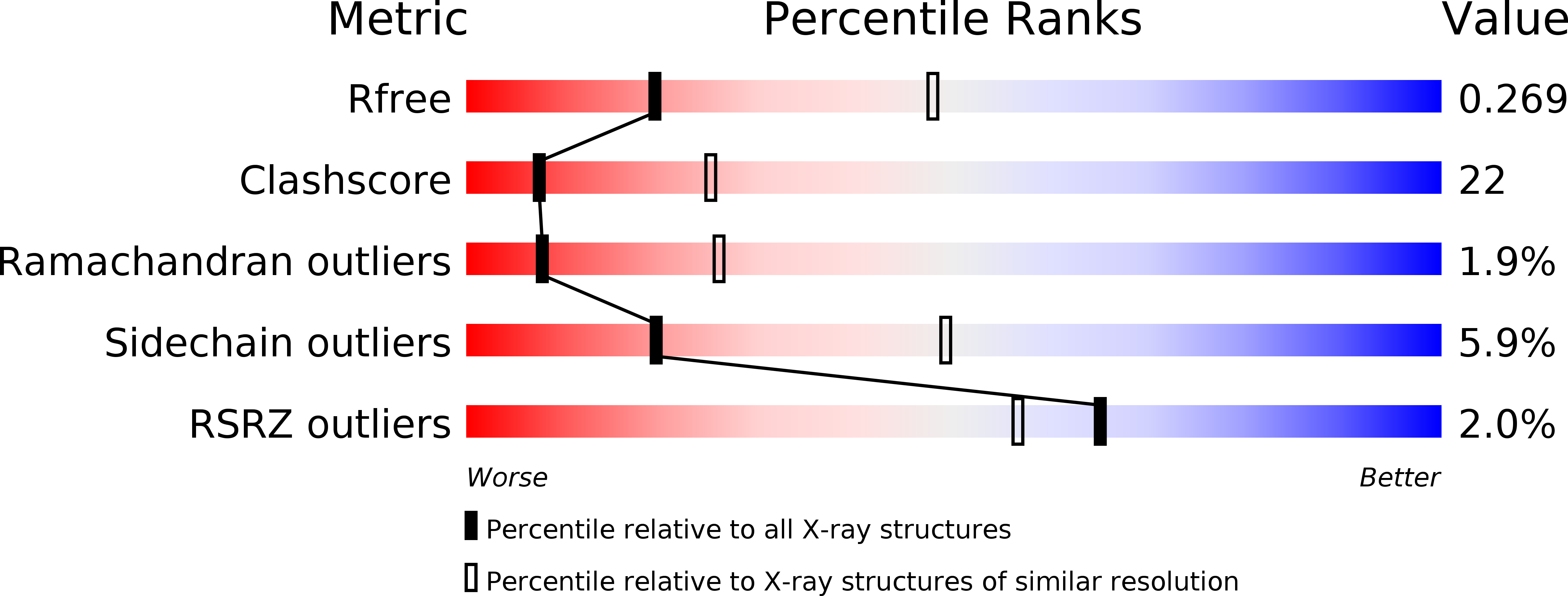
Resolution:
2.80 Å
R-Value Free:
0.27
R-Value Work:
0.22
Space Group:
P 21 21 21
Macromolecular Entities
Structures with similar UniProt ID
Protein Blast
Polymer Type:polypeptide(L)
Molecule:Fibrinogen alpha/alpha-E chain
Gene (Uniprot):FGA
Chain IDs:A, D
Chain Length:66
Number of Molecules:2
Biological Source:Homo sapiens
Structures with similar UniProt ID
Protein Blast
Polymer Type:polypeptide(L)
Molecule:Fibrinogen beta chain
Gene (Uniprot):FGB
Chain IDs:B, E
Chain Length:313
Number of Molecules:2
Biological Source:Homo sapiens
Structures with similar UniProt ID
Protein Blast
Polymer Type:polypeptide(L)
Molecule:Fibrinogen gamma chain
Gene (Uniprot):FGG
Chain IDs:C, F
Chain Length:311
Number of Molecules:2
Biological Source:Homo sapiens
Modified Residue
Compound ID Chain ID Parent Comp ID Details 2D Image
ASN B ASN GLYCOSYLATION SITE
Primary Citation
2.8 A Crystal Structures of Recombinant Fibrinogen Fragment D with and without Two Peptide Ligands: GHRP Binding to the "b" Site Disrupts Its Nearby Calcium-binding Site.
Biochemistry 41 12124 12132 (2002)
PMID: 12356313 DOI: 10.1021/bi0261894

Abstact

We report two crystal structures, each at a resolution of 2.8 A, of recombinant human fibrinogen fragment D (rfD) in the absence and presence of peptide ligands. The bound ligands, Gly-Pro-Arg-Pro-amide and Gly-His-Arg-Pro-amide, mimic the interactions of the thrombin exposed polymerization sites, "A" and "B", respectively. This report is the first to describe the structure of fragment D in the presence of both peptide ligands. The structures reveal that recombinant fibrinogen is nearly identical to the plasma protein but with minor changes, like the addition of a proximal fucose to the carbohydrate linked to residue betaGln364, and slightly different relative positions of the beta- and gamma-modules. Of major interest in our structures is that a previously identified calcium site in plasma fibrinogen is absent when Gly-His-Arg-Pro-amide is bound. The peptide-dependent loss of this calcium site may have significant biological implications that are further discussed. These structures provide a foundation for the detailed structural analysis of variant recombinant fibrinogens that were used to identify critical functional residues within fragment D.

Legend

Protein

Chemical

Disease

Primary Citation of related structures
Feedback Form
Name
Email
Institute
Feedback