1LS3 image
Deposition Date 2002-05-16
Release Date 2003-02-04
Last Version Date 2024-04-03
Entry Detail
PDB ID:
1LS3
Keywords:
Title:
Crystal Structure of the Complex between Rabbit Cytosolic Serine Hydroxymethyltransferase and TriGlu-5-formyl-tetrahydrofolate
Biological Source:
Source Organism:
Host Organism:
Method Details:
Experimental Method:
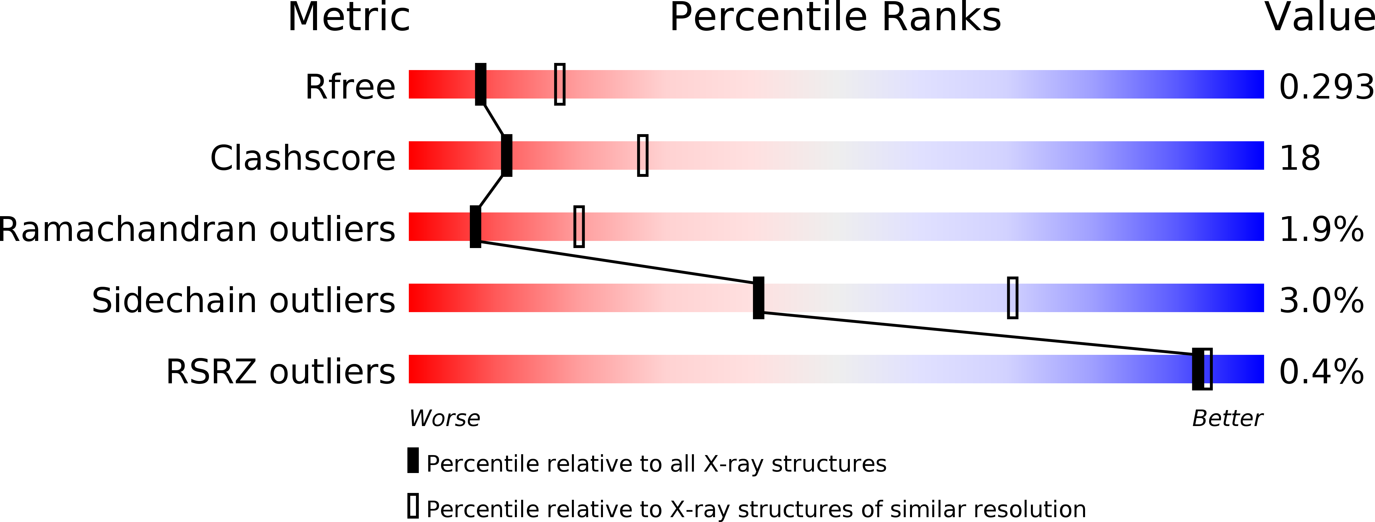
Resolution:
2.70 Å
R-Value Free:
0.29
R-Value Work:
0.22
R-Value Observed:
0.22
Space Group:
P 41
Macromolecular Entities
Structures with similar UniProt ID
Protein Blast
Polymer Type:polypeptide(L)
Molecule:Serine Hydroxymethyltransferase
Chain IDs:A, B, C, D
Chain Length:483
Number of Molecules:4
Biological Source:Oryctolagus cuniculus
Primary Citation
Location of the Pteroylpolyglutamate Binding Site on Rabbit Cytosolic Serine Hydroxymethyltransferase
J.Biol.Chem. 278 2645 2653 (2003)
PMID: 12438316 DOI: 10.1074/jbc.M210649200

Abstact

Serine hydroxymethyltransferase (SHMT; EC 2.1.2.1) catalyzes the reversible interconversion of serine and glycine with transfer of the serine side chain one-carbon group to tetrahydropteroylglutamate (H(4)PteGlu), and also the conversion of 5,10-methenyl-H(4)PteGlu to 5-formyl-H(4)PteGlu. In the cell, H(4)PteGlu carries a poly-gamma-glutamyl tail of at least 3 glutamyl residues that is required for physiological activity. This study combines solution binding and mutagenesis studies with crystallographic structure determination to identify the extended binding site for tetrahydropteroylpolyglutamate on rabbit cytosolic SHMT. Equilibrium binding and kinetic measurements of H(4)PteGlu(3) and H(4)PteGlu(5) with wild-type and Lys --> Gln or Glu site mutant homotetrameric rabbit cytosolic SHMTs identified lysine residues that contribute to the binding of the polyglutamate tail. The crystal structure of the enzyme in complex with 5-formyl-H(4)PteGlu(3) confirms the solution data and indicates that the conformation of the pteridine ring and its interactions with the enzyme differ slightly from those observed in complexes of the monoglutamate cofactor. The polyglutamate chain, which does not contribute to catalysis, exists in multiple conformations in each of the two occupied binding sites and appears to be bound by the electrostatic field created by the cationic residues, with only limited interactions with specific individual residues.

Legend

Protein

Chemical

Disease

Primary Citation of related structures