
Deposition Date
2002-05-16
Release Date
2002-09-04
Last Version Date
2024-02-14
Entry Detail
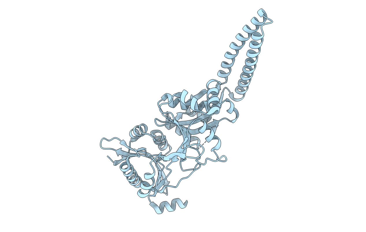
PDB ID:
1LRZ
Keywords:
Title:
x-ray crystal structure of staphylococcus aureus femA
Biological Source:
Source Organism(s):
Staphylococcus aureus (Taxon ID: 1280)
Expression System(s):
Method Details:
Experimental Method:
Resolution:
2.10 Å
R-Value Free:
0.21
R-Value Work:
0.18
R-Value Observed:
0.18
Space Group:
P 21 21 21
