1LRH image
Deposition Date 2002-05-15
Release Date 2002-06-19
Last Version Date 2024-11-13
Entry Detail
PDB ID:
1LRH
Keywords:
Title:
Crystal structure of auxin-binding protein 1 in complex with 1-naphthalene acetic acid
Biological Source:
Source Organism:
Zea mays (Taxon ID: 4577)
Host Organism:
Method Details:
Experimental Method:
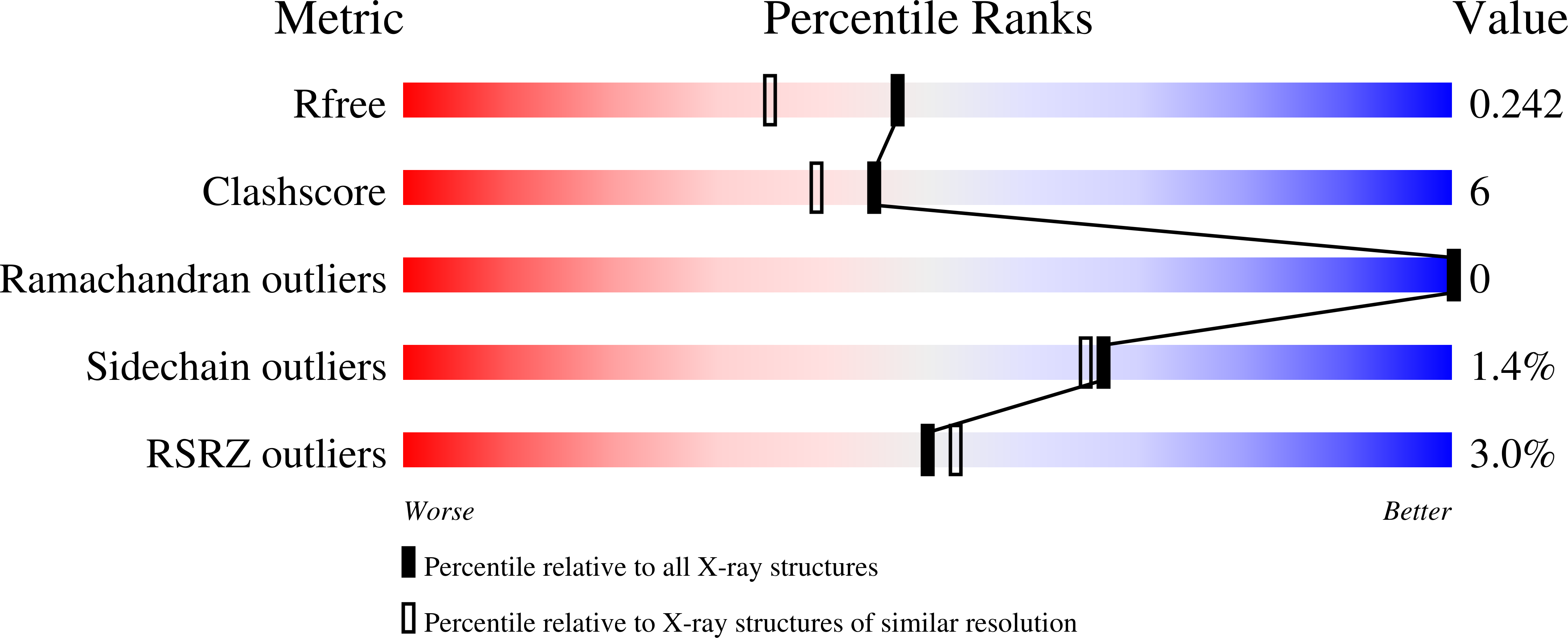
Resolution:
1.90 Å
R-Value Free:
0.24
R-Value Work:
0.2
R-Value Observed:
0.2
Space Group:
P 1 21 1
Macromolecular Entities
Structures with similar UniProt ID
Protein Blast
Polymer Type:polypeptide(L)
Molecule:auxin-binding protein 1
Gene (Uniprot):ABP1
Mutagens:D161E/E162Q
Chain IDs:A, B, C, D
Chain Length:163
Number of Molecules:4
Biological Source:Zea mays
Modified Residue
Compound ID Chain ID Parent Comp ID Details 2D Image
ASN A ASN GLYCOSYLATION SITE
Primary Citation
Crystal structure of auxin-binding protein 1 in complex with auxin.
EMBO J. 21 2877 2885 (2002)
PMID: 12065401 DOI: 10.1093/emboj/cdf291

Abstact

The structure of auxin-binding protein 1 (ABP1) from maize has been determined at 1.9 A resolution, revealing its auxin-binding site. The structure confirms that ABP1 belongs to the ancient and functionally diverse germin/seed storage 7S protein superfamily. The binding pocket of ABP1 is predominantly hydrophobic with a metal ion deep inside the pocket coordinated by three histidines and a glutamate. Auxin binds within this pocket, with its carboxylate binding the zinc and its aromatic ring binding hydrophobic residues including Trp151. There is a single disulfide between Cys2 and Cys155. No conformational rearrangement of ABP1 was observed when auxin bound to the protein in the crystal, but examination of the structure reveals a possible mechanism of signal transduction.

Legend

Protein

Chemical

Disease

Primary Citation of related structures