1LQM image
Deposition Date 2002-05-10
Release Date 2002-11-10
Last Version Date 2024-02-14
Entry Detail
PDB ID:
1LQM
Title:
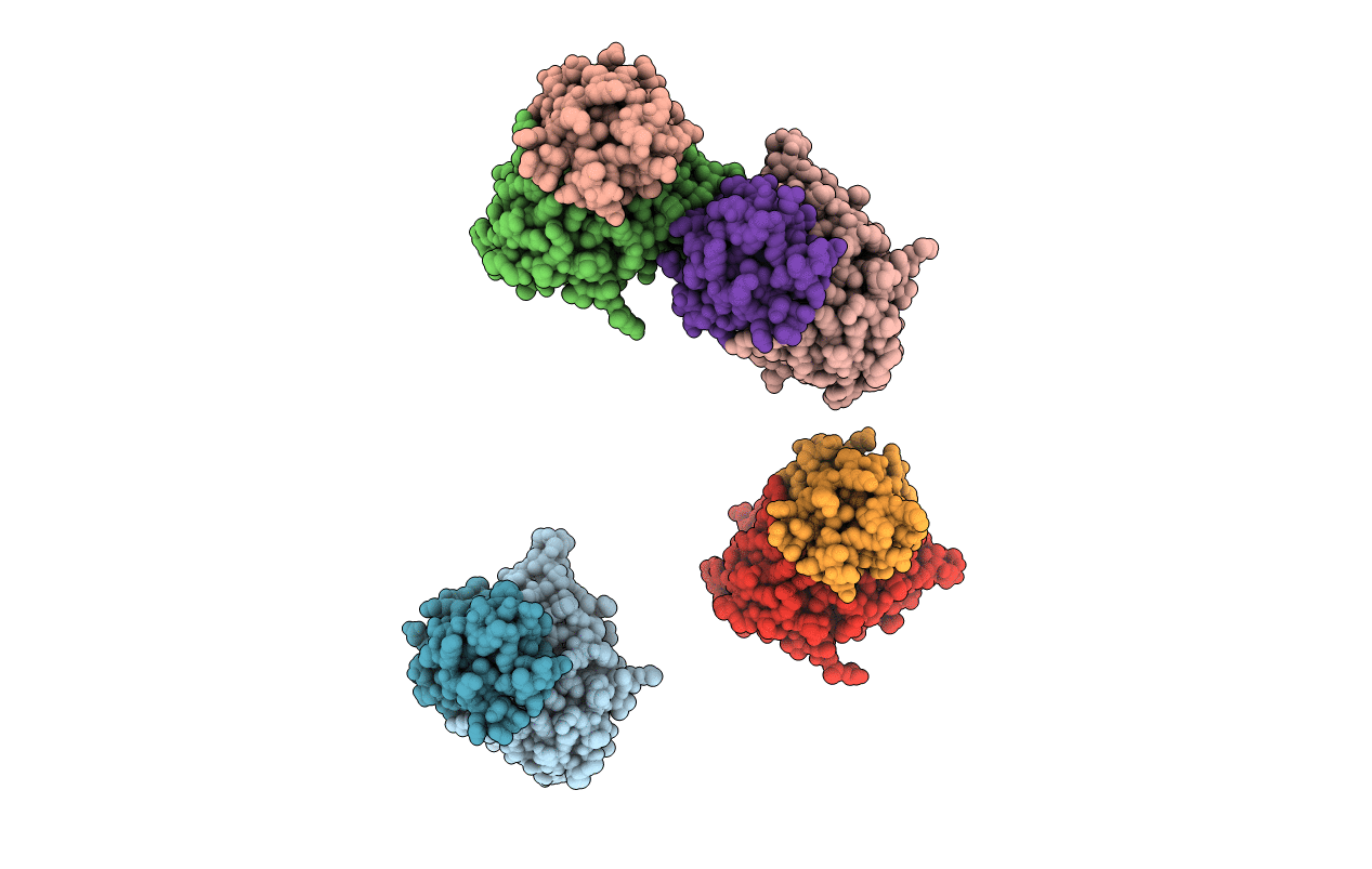
ESCHERICHIA COLI URACIL-DNA GLYCOSYLASE COMPLEX WITH URACIL-DNA GLYCOSYLASE INHIBITOR PROTEIN
Biological Source:
Source Organism:
Host Organism:
Method Details:
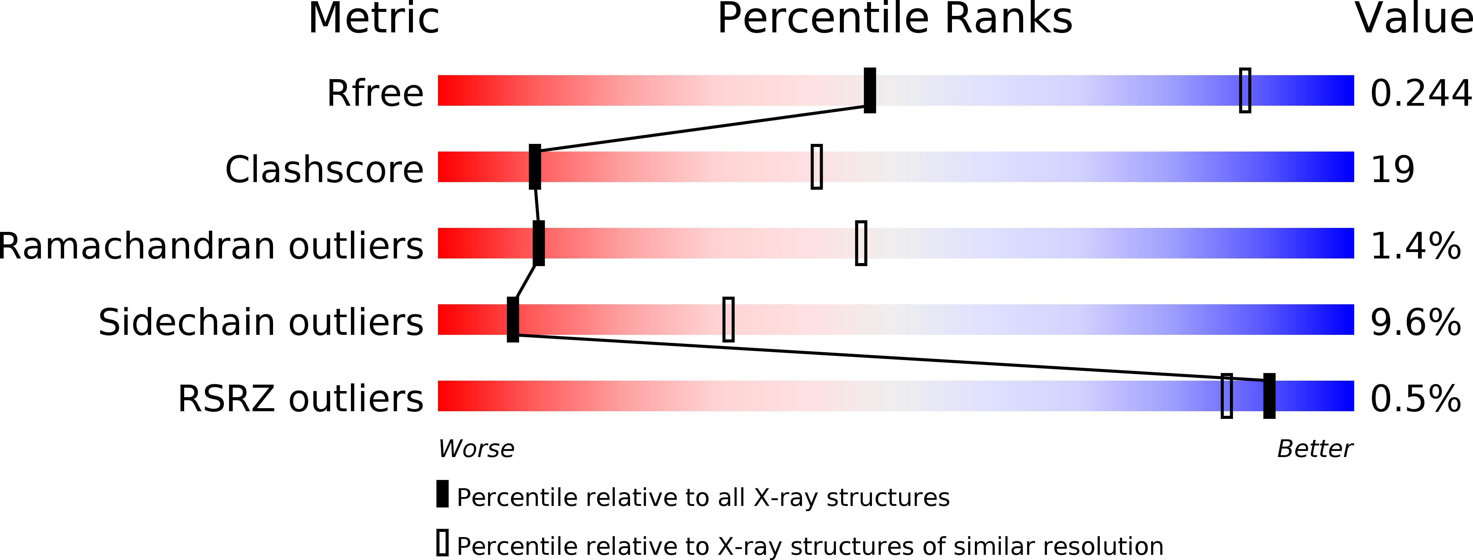
Experimental Method:
Resolution:
3.20 Å
R-Value Free:
0.26
R-Value Work:
0.18
R-Value Observed:
0.18
Space Group:
P 21 21 2
Macromolecular Entities
Polymer Type:polypeptide(L)
Molecule:URACIL-DNA GLYCOSYLASE
Gene (Uniprot):ung
Chain IDs:A, C, E, G
Chain Length:229
Number of Molecules:4
Biological Source:Escherichia coli
Polymer Type:polypeptide(L)
Molecule:URACIL-DNA GLYCOSYLASE INHIBITOR
Gene (Uniprot):UGI
Chain IDs:B, D, F, H
Chain Length:84
Number of Molecules:4
Biological Source:Bacillus phage PBS2
Primary Citation
Domain closure and action of uracil DNA glycosylase (UDG): structures of new crystal forms containing the Escherichia coli enzyme and a comparative study of the known structures involving UDG.
Acta Crystallogr.,Sect.D 58 1269 1276 (2002)
PMID: 12136137 DOI: 10.1107/S0907444902009599

Abstact

The structures of a new crystal form of free Escherichia coli uracil DNA glycosylase (UDG), containing four molecules in the asymmetric unit, and two forms of its complex with the proteinaceous inhibitor Ugi, containing two and four crystallographically independent complexes, have been determined. A comparison of these structures and the already known crystal structures containing UDG shows that the enzyme can be considered to be made up of two independently moving structural entities or domains. A detailed study of free and DNA-bound human enzyme strengthens this conclusion. The domains close upon binding to uracil-containing DNA, whereas they do not appear to do so upon binding to Ugi. The comparative study also shows that the mobility of the molecule involves the rigid-body movement of the domains superposed on flexibility within domains.

Legend

Protein

Chemical

Disease

Primary Citation of related structures