1LPG image
Entry Detail
PDB ID:
1LPG
Keywords:
Title:
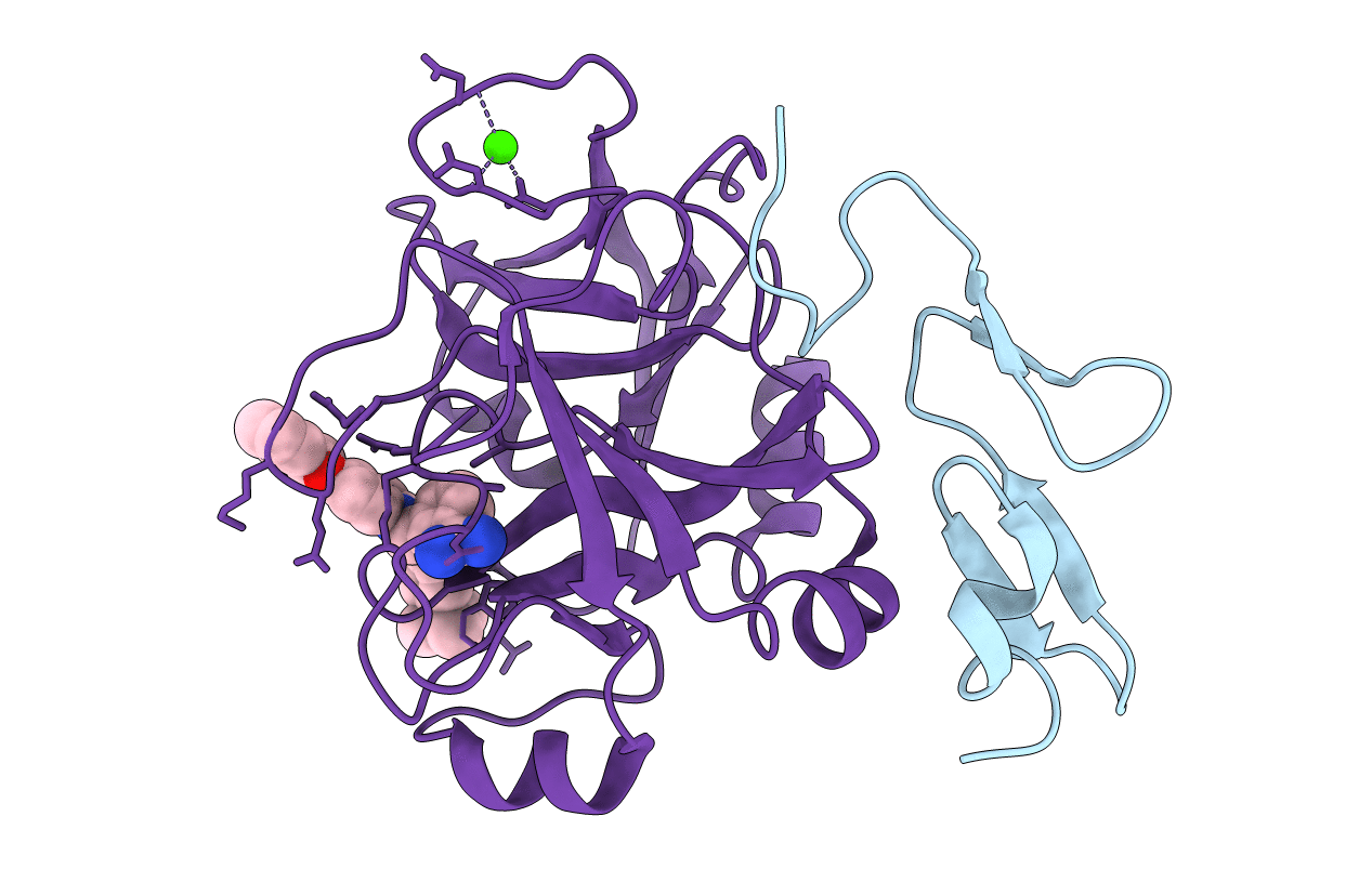
CRYSTAL STRUCTURE OF FXA IN COMPLEX WITH 79.
Biological Source:
Source Organism:
PDB Version:
Deposition Date:
2002-05-08
Release Date:
2003-05-08
Method Details:
Experimental Method:
Resolution:
2.00 Å
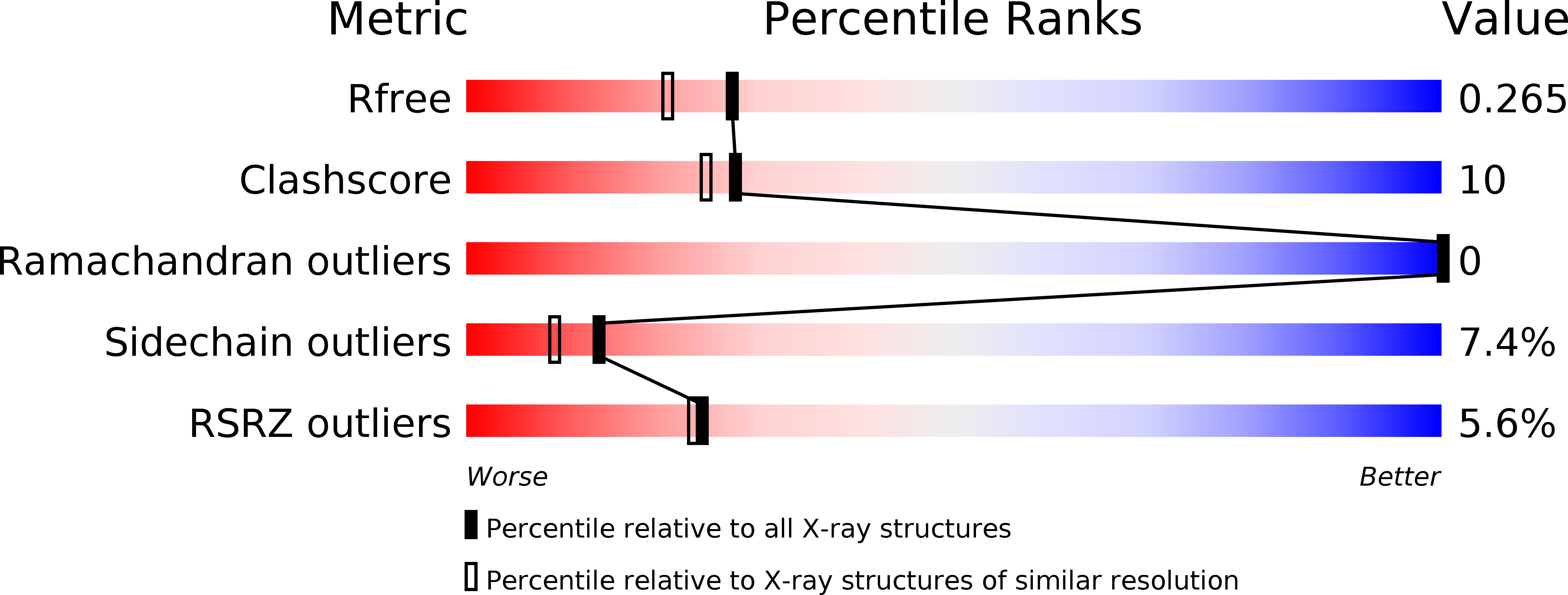
R-Value Free:
0.28
R-Value Work:
0.16
R-Value Observed:
0.20
Space Group:
P 21 21 21
Macromolecular Entities
Polymer Type:polypeptide(L)
Description:Blood coagulation factor Xa
Chain IDs:A
Chain Length:134
Number of Molecules:1
Biological Source:Homo sapiens
Polymer Type:polypeptide(L)
Description:Blood coagulation factor Xa
Chain IDs:B
Chain Length:254
Number of Molecules:1
Biological Source:Homo sapiens
Primary Citation
Design and Quantitative Structure-Activity relationship of 3-amidinobenzyl-1H-indole-2-carboxamides as potent, nonchiral, and selective inhibitors of blood coagulation factor Xa.
J.Med.Chem. 45 2749 2769 (2002)
PMID: 12061878 DOI: 10.1021/jm0111346

Abstact

A series of 138 nonchiral 3-amidinobenzyl-1H-indole-2-carboxamides and analogues as inhibitors of the blood coagulation enzyme factor Xa (fXa) were designed, synthesized, and investigated by X-ray structure analysis and 3D quantitative structure-activity relationship (QSAR) studies (CoMFA, CoMSIA) in order to identify important protein-ligand interactions responsible for biological affinity and selectivity. Several compounds from this series are highly potent and selective inhibitors of this important enzyme linking extrinsic and intrinsic coagulation pathways. To rationalize biological affinity and to provide guidelines for further design, all compounds were docked into the factor Xa binding site. Those docking studies were based on X-ray structures of factor Xa in complex with literature-known inhibitors. It was possible to validate those binding modes by four X-ray crystal structures of representative ligands in factor Xa, while one ligand was additionally crystallized in trypsin to rationalize requirements for selective factor Xa inhibition. The 3D-QSAR models based on a superposition rule derived from these docking studies were validated using conventional and cross-validated r(2) values using the leave-one-out method and repeated analyses using two randomly chosen cross-validation groups plus randomization of biological activities. This led to consistent and highly predictive 3D-QSAR models with good correlation coefficients for both CoMFA and CoMSIA, which were found to correspond to experimentally determined factor Xa binding site topology in terms of steric, electrostatic, and hydrophobic complementarity. Subsets selected as smaller training sets using 2D fingerprints and maximum dissimilarity methods resulted in 3D-QSAR models with remarkable correlation coefficients and a high predictive power. The final quantitative SAR information agrees with all experimental data for the binding topology and thus provides reasonable activity predictions for novel factor Xa inhibitors.

Legend

Protein

Chemical

Disease

Primary Citation of related structures