1LOP image
Deposition Date 1996-06-17
Release Date 1996-12-23
Last Version Date 2025-03-26
Entry Detail
PDB ID:
1LOP
Title:
CYCLOPHILIN A COMPLEXED WITH SUCCINYL-ALA-PRO-ALA-P-NITROANILIDE
Biological Source:
Source Organism:
Escherichia coli (Taxon ID: 562)
Method Details:
Experimental Method:
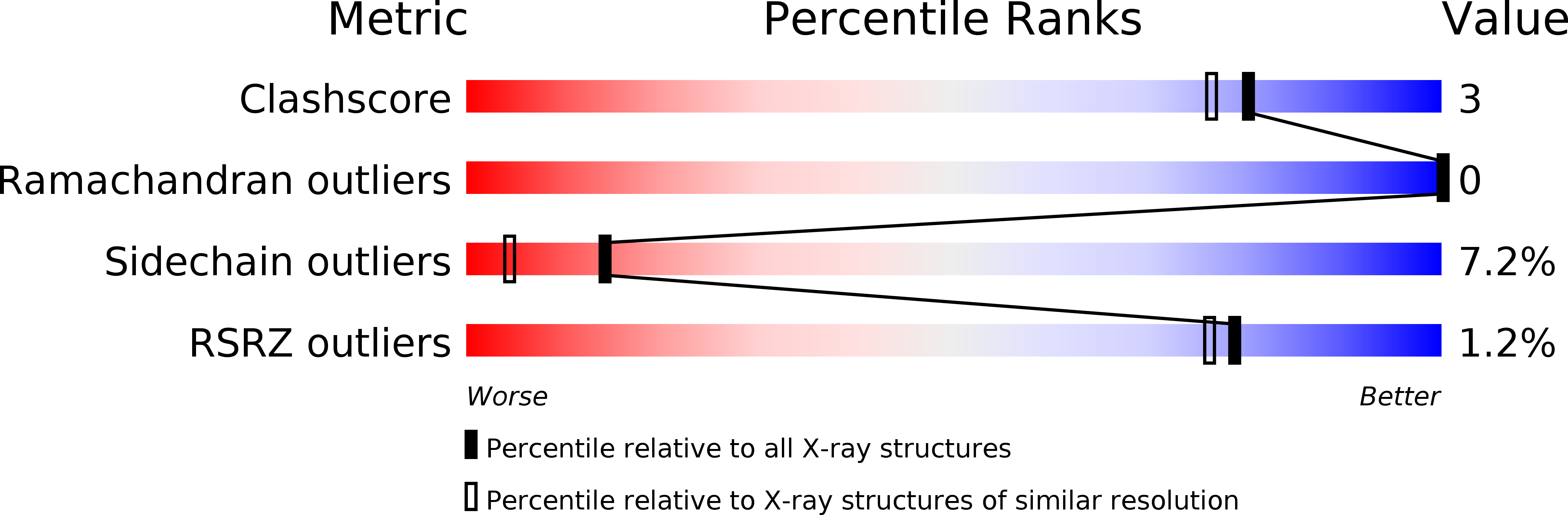
Resolution:
1.80 Å
R-Value Work:
0.17
R-Value Observed:
0.17
Space Group:
P 21 21 21
Macromolecular Entities
Structures with similar UniProt ID
Protein Blast
Polymer Type:polypeptide(L)
Molecule:CYCLOPHILIN A
Gene (Uniprot):ppiB
Chain IDs:A
Chain Length:164
Number of Molecules:1
Biological Source:Escherichia coli
Peptide-like Molecules
PRD_000400
Primary Citation
The substrate-binding site in Escherichia coli cyclophilin A preferably recognizes a cis-proline isomer or a highly distorted form of the trans isomer.
J.Mol.Biol. 256 897 908 (1996)
PMID: 8601841 DOI: 10.1006/jmbi.1996.0136

Abstact

The three-dimensional structure of Escherichia coli cytosolic cyclophilin A (CyPA) complexed with a tripeptide (succinyl-Ala-Pro-Ala-p-nitroanilide) was refined at 1.8 A resolution by the multiple isomorphous replacement method to a crystallographic R-factor of 17.6%. As in human CyPA, the peptide binding site in E. coli enzyme is in a cleft created on the surface of the upper sheet of two orthogonal beta-sheets. In this cleft, the walls of the hydrophobic pocket are formed by the side-chains of five non-polar residues, Phe48, Met49, Phe107, Leu108, and Try120, with Phe99 at the bottom. When the cis isomer of the tripeptide binds to the enzyme, a cis-proline ring is inserted into the hydrophobic pocket. Since the binding pocket of CyPAs are largely hydrophobic, the cis isomer of a peptide can be bound more firmly than the trans isomer. Distortion of the trans isomer could lead to better binding, but at an energetic cost of the distortion energy. At the periphery of the upper beta-sheet in E. coli CyPA, conformations of loops L1, L3, and L4 and the segment connecting alpha1 and beta3 with deletions or insertions against human CyPA differ significantly from those in human CyPA. The refined model also shows that steric hindrance to attachment of cyclosporin A (CsA) prevents E. coli CyPA forming a complex with CsA. Thus, the extra amino acid residue of E. coli CyPA, polar Gln89, lies along the pathway to the hydrophobic pocket of CyPA and seems to prevent the access hydrophobic part of CsA to the cleft of CyPA.

Legend

Protein

Chemical

Disease

Primary Citation of related structures
Feedback Form
Name
Email
Institute
Feedback