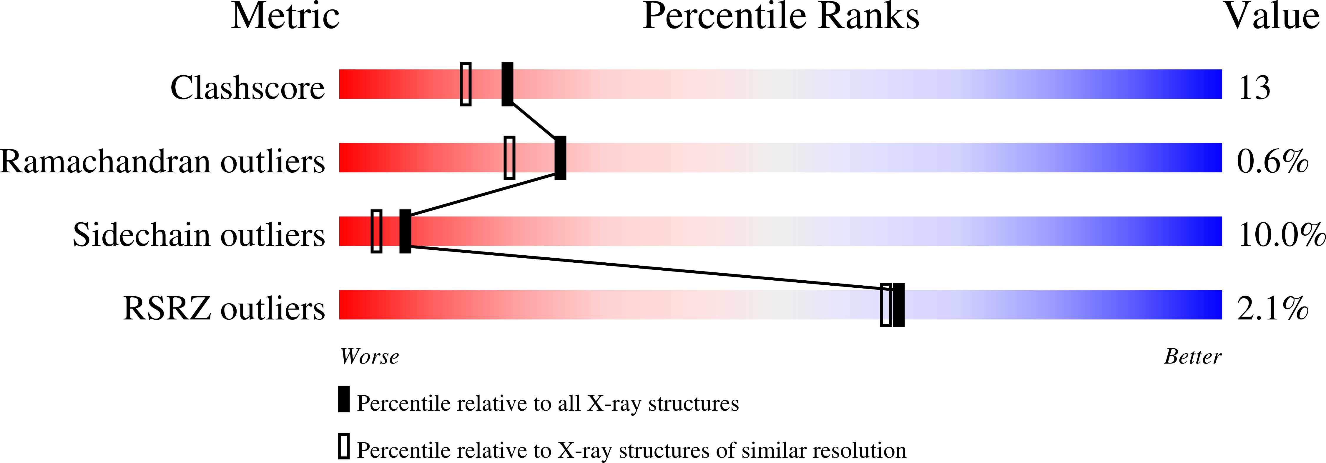
1LOH image
Deposition Date 2002-05-06
Release Date 2002-08-07
Last Version Date 2024-02-14
Entry Detail
PDB ID:
1LOH
Keywords:
Title:
Streptococcus pneumoniae Hyaluronate Lyase in Complex with Hexasaccharide Hyaluronan Substrate
Biological Source:
Source Organism:
Host Organism:
Method Details:
Experimental Method:
Resolution:
2.00 Å
R-Value Free:
0.32
R-Value Work:
0.22
R-Value Observed:
0.22
Space Group:
P 21 21 21
Macromolecular Entities
Polymer Type:polypeptide(L)
Molecule:Hyaluronate Lyase
Mutations:Y408F
Chain IDs:A
Chain Length:721
Number of Molecules:1
Biological Source:Streptococcus pneumoniae
Primary Citation
Mechanism of hyaluronan degradation by Streptococcus pneumoniae hyaluronate lyase. Structures of complexes with the substrate.
J.Biol.Chem. 277 28287 28297 (2002)
PMID: 11991948 DOI: 10.1074/jbc.M112009200

Abstact

Hyaluronate lyase enzymes degrade hyaluronan, the main polysaccharide component of the host connective tissues, predominantly into unsaturated disaccharide units, thereby destroying the normal connective tissue structure and exposing the tissue cells to various endo- and exogenous factors, including bacterial toxins. The crystal structures of Streptococcus pneumoniae hyaluronate lyase with tetra- and hexasaccharide hyaluronan substrates bound in the active site were determined at 1.52- and 2.0-A resolution, respectively. Hexasaccharide is the longest substrate segment that binds entirely within the active site of these enzymes. The enzyme residues responsible for substrate binding, positioning, catalysis, and product release were thereby identified and their specific roles characterized. The involvement of three residues in catalysis, Asn(349), His(399), and Tyr(408), is confirmed, and the details of proton acceptance and donation within the catalytic machinery are described. The mechanism of processivity of the enzyme is analyzed. The flexibility (allosteric) behavior of the enzyme may be understood in terms of the results of flexibility analysis of this protein, which identified two modes of motion that are also proposed to be involved in the hyaluronan degradation process. The first motion describes an opening and closing of the catalytic cleft located between the alpha- and beta-domains. The second motion demonstrates the mobility of a binding cleft, which may facilitate the binding of the negatively charged hyaluronan to the enzyme.

Legend

Protein

Chemical

Disease

Primary Citation of related structures