Deposition Date
1993-01-27
Release Date
1994-04-30
Last Version Date
2024-02-14
Entry Detail
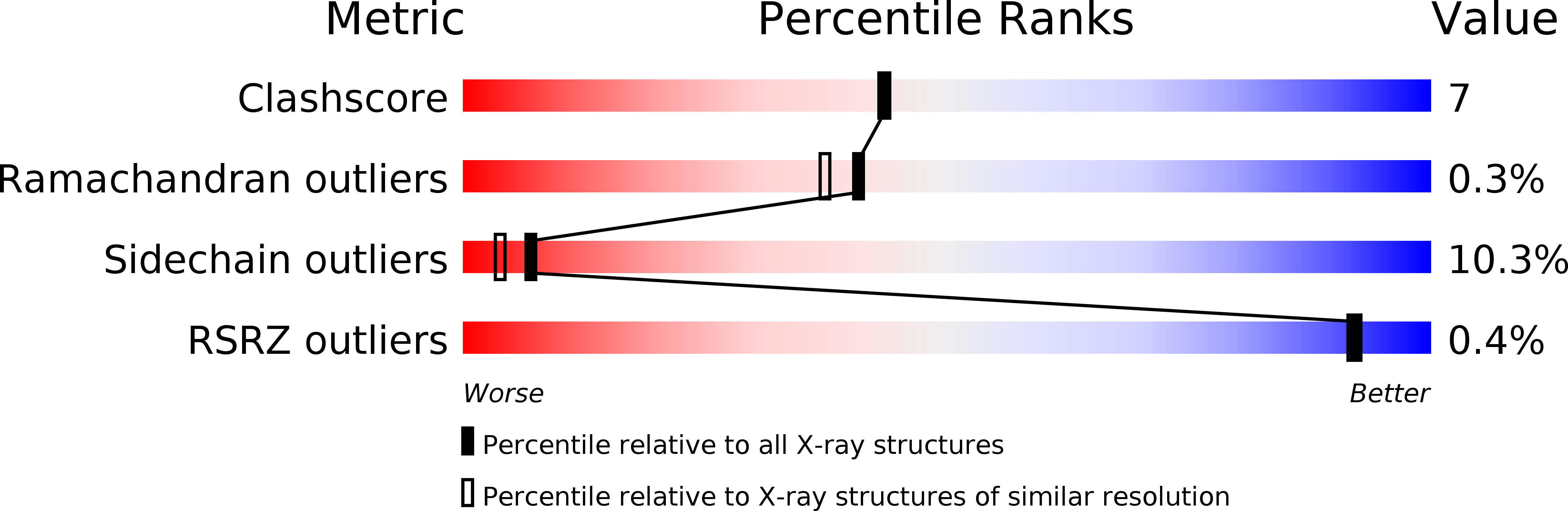
PDB ID:
1LOB
Keywords:
Title:
THREE-DIMENSIONAL STRUCTURES OF COMPLEXES OF LATHYRUS OCHRUS ISOLECTIN I WITH GLUCOSE AND MANNOSE: FINE SPECIFICITY OF THE MONOSACCHARIDE-BINDING SITE
Biological Source:
Source Organism(s):
Lathyrus ochrus (Taxon ID: 3858)
Method Details:
Experimental Method:
Resolution:
2.00 Å
R-Value Work:
0.18
R-Value Observed:
0.18
Space Group:
P 1 21 1