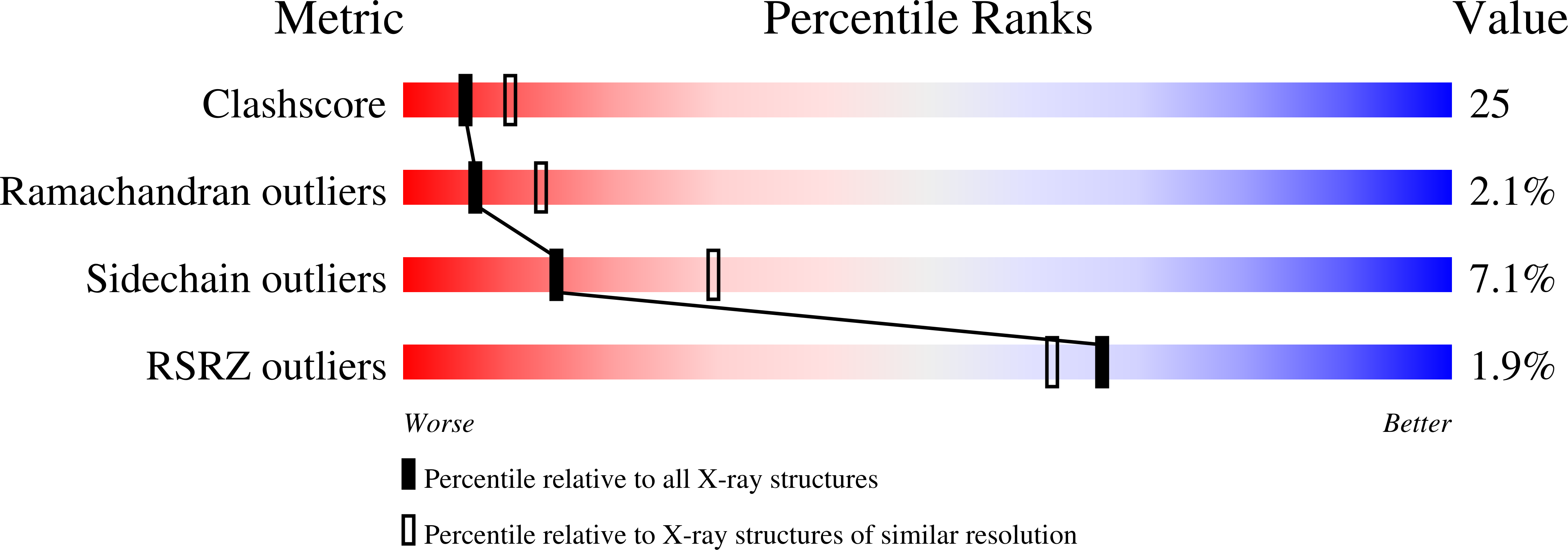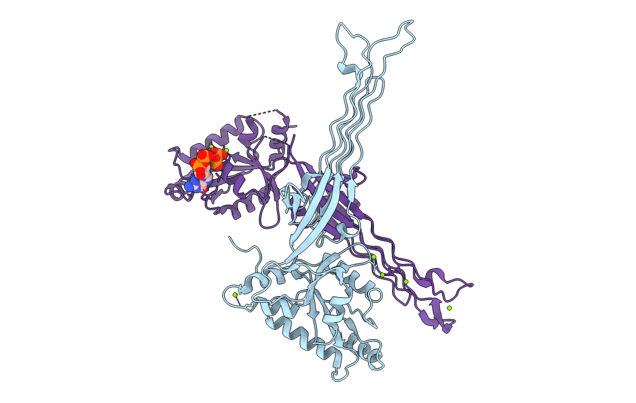
1LNZ image
Deposition Date 2002-05-04
Release Date 2002-09-16
Last Version Date 2024-11-20
Entry Detail
PDB ID:
1LNZ
Keywords:
Title:
Structure of the Obg GTP-binding protein
Biological Source:
Source Organism:
Host Organism:
Method Details:
Experimental Method:
Resolution:
2.60 Å
R-Value Free:
0.29
R-Value Work:
0.20
R-Value Observed:
0.20
Space Group:
P 21 21 21
Macromolecular Entities
Polymer Type:polypeptide(L)
Molecule:SPO0B-associated GTP-binding protein
Gene (Uniprot):obg
Mutations:selenomethionine substituted
Chain IDs:A, B
Chain Length:342
Number of Molecules:2
Biological Source:Bacillus subtilis
Modified Residue
Compound ID Chain ID Parent Comp ID Details 2D Image
MSE A MET SELENOMETHIONINE
Primary Citation
Structural and biochemical analysis of the Obg GTP binding protein
Structure 10 1581 1592 (2002)
PMID: 12429099 DOI: 10.1016/S0969-2126(02)00882-1

Abstact

The Obg nucleotide binding protein family has been implicated in stress response, chromosome partitioning, replication initiation, mycelium development, and sporulation. Obg proteins are among a large group of GTP binding proteins conserved from bacteria to man. Members of the family contain two equally and highly conserved domains, a C-terminal GTP binding domain and an N-terminal glycine-rich domain. Structural analysis of Bacillus subtilis Obg revealed respective domain architectures and how they are coupled through the putative switch elements of the C-terminal GTPase domain in apo and nucleotide-bound configurations. Biochemical analysis of bacterial and human Obg proteins combined with the structural observation of the ppGpp nucleotide within the Obg active sight suggest a potential role for ppGpp modulation of Obg function in B. subtilis.

Legend

Protein

Chemical

Disease

Primary Citation of related structures