1LN0 image
Deposition Date 2002-05-02
Release Date 2002-10-30
Last Version Date 2024-10-30
Entry Detail
PDB ID:
1LN0
Keywords:
Title:
Structure of the Catalytic Domain of Homing Endonuclease I-TevI
Biological Source:
Source Organism:
Host Organism:
Method Details:
Experimental Method:
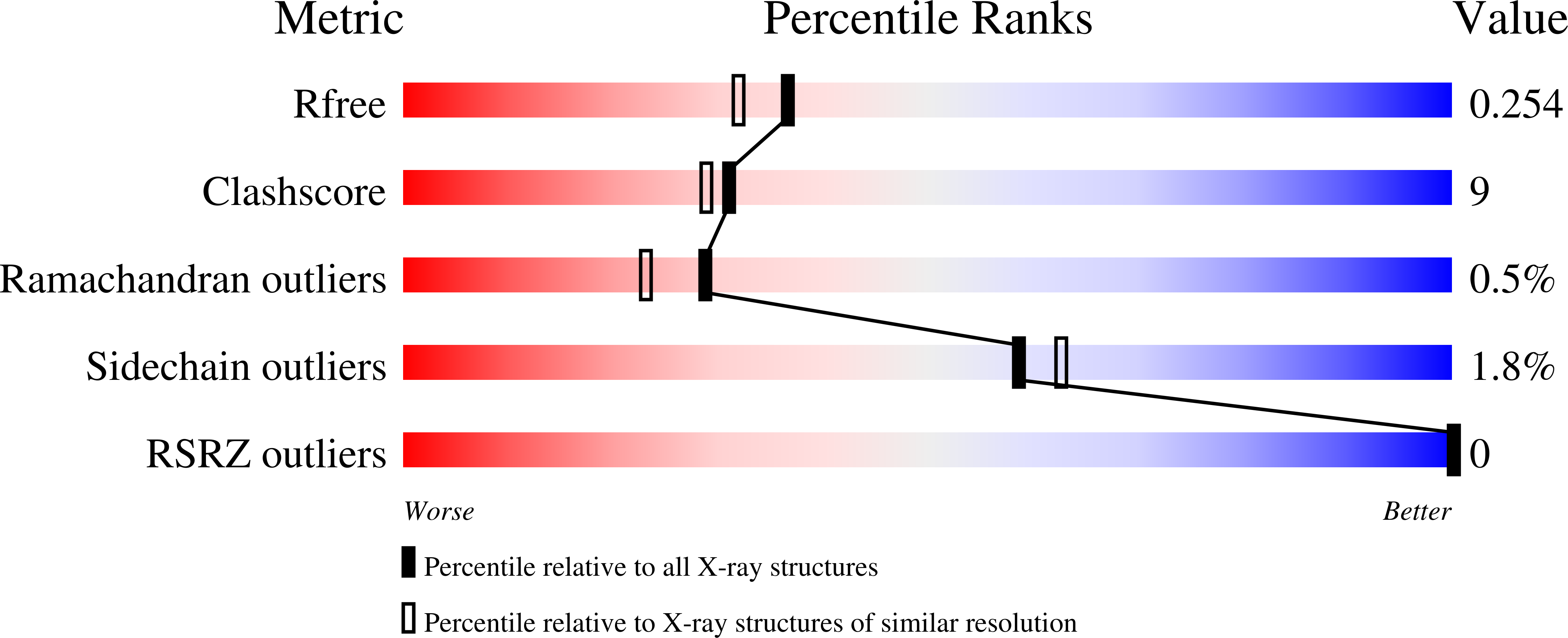
Resolution:
2.00 Å
R-Value Free:
0.25
R-Value Work:
0.20
R-Value Observed:
0.20
Space Group:
P 21 21 2
Macromolecular Entities
Polymer Type:polypeptide(L)
Molecule:intron-associated endonuclease 1
Gene (Uniprot):ITEVIR
Mutations:R27A
Chain IDs:A, B
Chain Length:97
Number of Molecules:2
Biological Source:Enterobacteria phage T4
Ligand Molecules
Primary Citation
Catalytic domain structure and hypothesis for function of GIY-YIG intron endonuclease I-TevI.
Nat.Struct.Biol. 9 806 811 (2002)
PMID: 12379841

Abstact

I-TevI, a member of the GIY-YIG family of homing endonucleases, consists of an N-terminal catalytic domain and a C-terminal DNA-binding domain joined by a flexible linker. The GIY-YIG motif is in the N-terminal domain of I-TevI, which corresponds to a phylogenetically widespread catalytic cartridge that is often associated with mobile genetic elements. The crystal structure of the catalytic domain of I-TevI, the first of any GIY-YIG endonuclease, reveals a novel alpha/beta-fold with a central three-stranded antiparallel beta-sheet flanked by three helices. The most conserved and putative catalytic residues are located on a shallow, concave surface and include a metal coordination site. Similarities in the three-dimensional arrangement of the catalytically important residues and the cation-binding site with those of the His-Cys box endonuclease I-PpoI suggest the possibility of mechanistic relationships among these different families of homing endonucleases despite completely different folds.

Legend

Protein

Chemical

Disease

Primary Citation of related structures