1LI4 image
Deposition Date 2002-04-17
Release Date 2003-05-20
Last Version Date 2023-08-16
Entry Detail
PDB ID:
1LI4
Keywords:
Title:
Human S-adenosylhomocysteine hydrolase complexed with neplanocin
Biological Source:
Source Organism:
Homo sapiens (Taxon ID: 9606)
Host Organism:
Method Details:
Experimental Method:
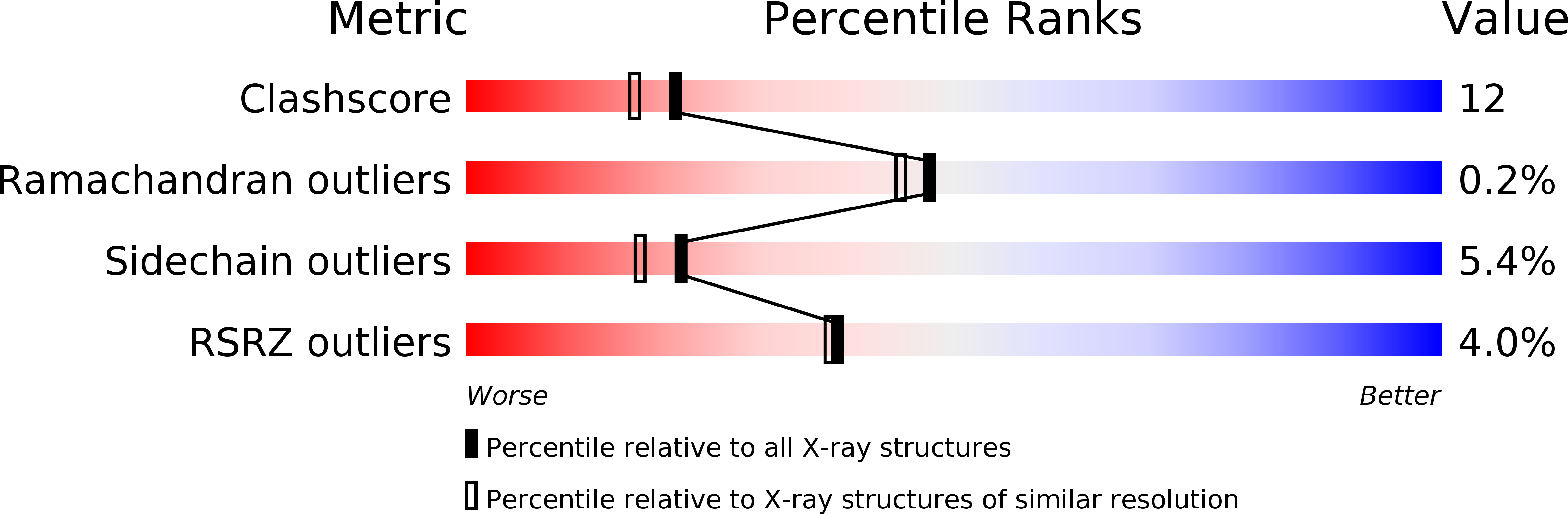
Resolution:
2.01 Å
R-Value Free:
0.23
R-Value Work:
0.19
R-Value Observed:
0.19
Space Group:
F 2 2 2
Macromolecular Entities
Structures with similar UniProt ID
Protein Blast
Polymer Type:polypeptide(L)
Molecule:S-adenosylhomocysteine hydrolase
Gene (Uniprot):AHCY
Chain IDs:A
Chain Length:432
Number of Molecules:1
Biological Source:Homo sapiens
Primary Citation
Catalytic strategy of S-adenosyl-L-homocysteine hydrolase: Transition-state stabilization and the avoidance of abortive reactions
Biochemistry 42 1900 1909 (2003)
PMID: 12590576 DOI: 10.1021/bi0262350

Abstact

S-Adenosylhomocysteine hydrolase (AdoHcy hydrolase) crystallizes from solutions containing the intermediate analogue neplanocin A with the analogue bound in its 3'-keto form at the active sites of all of its four subunits and the four tightly bound cofactors in their reduced (NADH) state. The enzyme is in the closed conformation, which corresponds to the structure in which the catalytic chemistry occurs. Examination of the structure in the light of available, very detailed kinetic studies [Porter, D. J., Boyd, F. L. (1991) J. Biol. Chem. 266, 21616-21625. Porter, D. J., Boyd, F. L. (1992) J. Biol. Chem. 267, 3205-3213. Porter, D. J. (1998) J. Biol. Chem. 268, 66-73] suggests elements of the catalytic strategy of AdoHcy hydrolase for acceleration of the reversible conversion of AdoHcy to adenosine (Ado) and homocysteine (Hcy). The enzyme, each subunit of which possesses a substrate-binding domain that in the absence of substrate is in rapid motion relative to the tetrameric core of the enzyme, first binds substrate and ceases motion. Probably concurrently with oxidation of the substrate to its 3'-keto form, the closed active site is "sealed off" from the environment, as indicated by a large (10(8)(-)(9)-fold) reduction in the rate of departure of ligands, a feature that prevents exposure of the labile 3'-keto intermediates to the aqueous environment. Elimination of the 5'-substituent (Hcy in the hydrolytic direction, water in the synthetic direction) generates the central intermediate 4',5'-didehydro-5'-deoxy-3'-ketoadenosine. Abortive 3'-reduction of the central intermediate is prevented by a temporary suspension of all or part of the redox catalytic power of the enzyme during the existence of the central intermediate. The abortive reduction is 10(4)-fold slower than the productive reductions at the ends of the catalytic cycle and has a rate constant similar to those of nonenzymic intramolecular model reactions. The mechanism for suspending the redox catalytic power appears to be a conformationally induced increase in the distance across which hydride transfer must occur between cofactor and substrate, the responsible conformational change again being that which "seals" the active site. The crystal structure reveals a well-defined chain of three water molecules leading from the active site to the subunit surface, which may serve as a relay for proton exchange between solvent and active site in the closed form of the enzyme, permitting maintenance of active-site functional groups in catalytically suitable protonation states.

Legend

Protein

Chemical

Disease

Primary Citation of related structures