1LHN image
Deposition Date 2002-04-17
Release Date 2002-10-23
Last Version Date 2024-10-30
Entry Detail
PDB ID:
1LHN
Title:
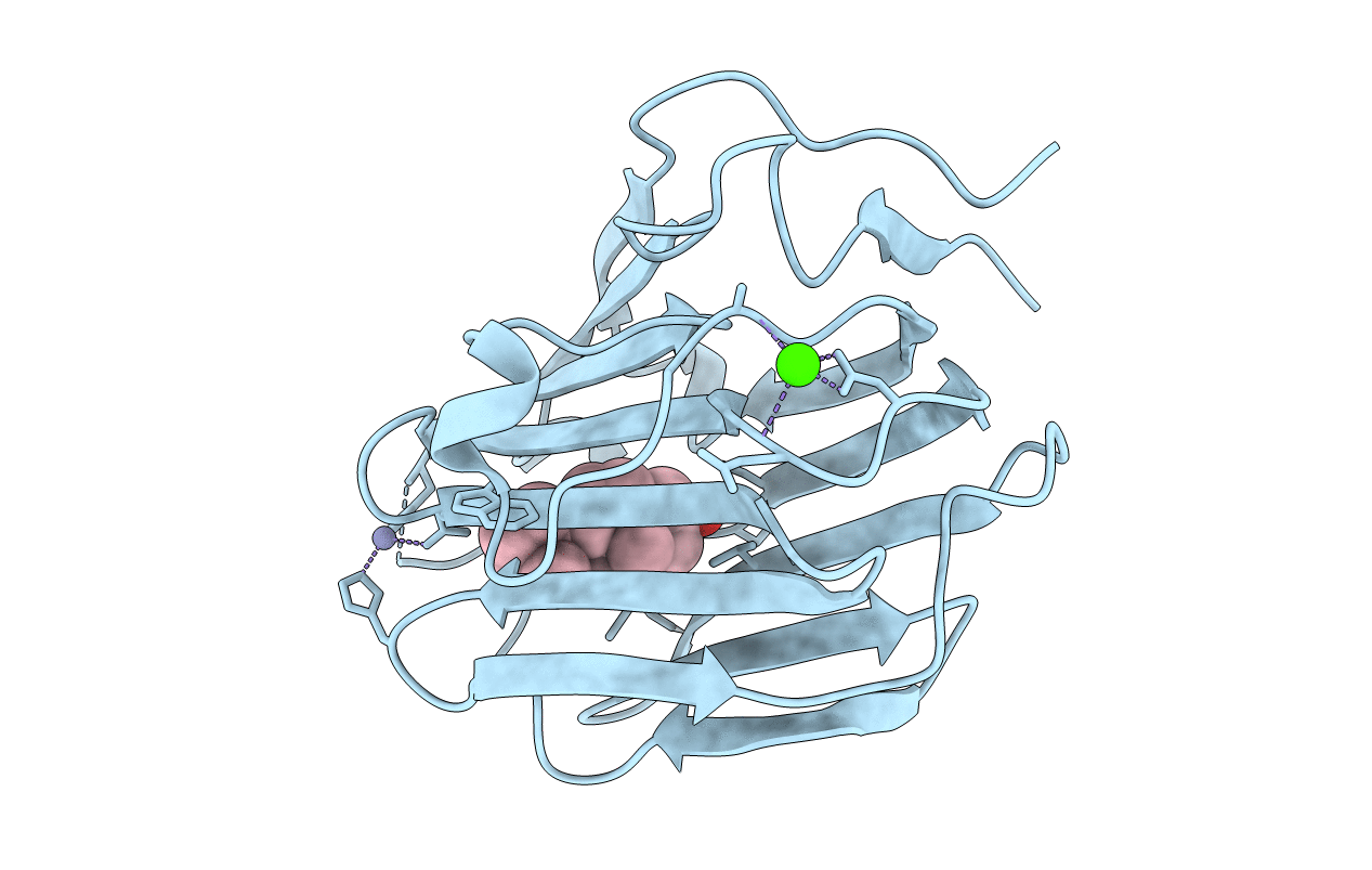
CRYSTAL STRUCTURE OF THE N-TERMINAL LG-DOMAIN OF SHBG IN COMPLEX WITH 5ALPHA-ANDROSTANE-3BETA,17ALPHA-DIOL
Biological Source:
Source Organism:
Homo sapiens (Taxon ID: 9606)
Host Organism:
Method Details:
Experimental Method:
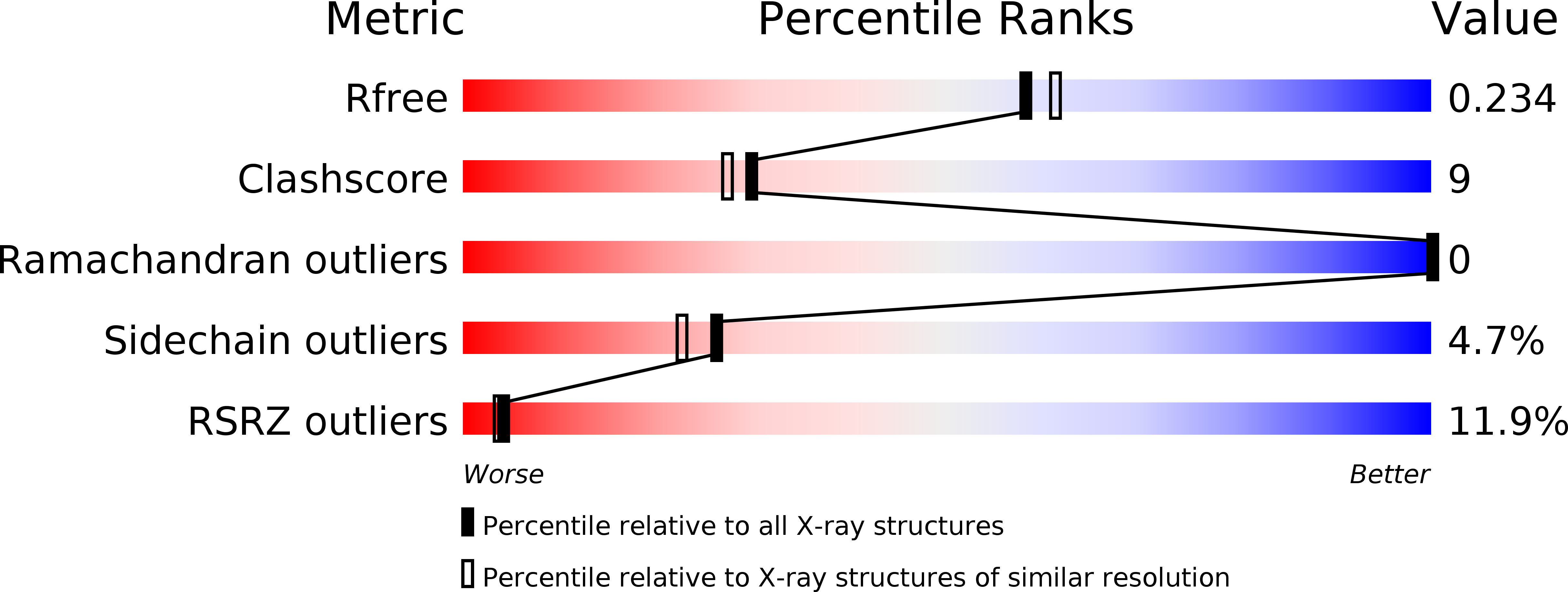
Resolution:
2.00 Å
R-Value Free:
0.23
R-Value Work:
0.19
Space Group:
H 3 2
Macromolecular Entities
Structures with similar UniProt ID
Protein Blast
Polymer Type:polypeptide(L)
Molecule:SEX HORMONE-BINDING GLOBULIN
Gene (Uniprot):SHBG
Chain IDs:A
Chain Length:189
Number of Molecules:1
Biological Source:Homo sapiens
Primary Citation
Steroid Ligands Bind Human Sex Hormone-binding Globulin in Specific Orientations and Produce Distinct Changes in Protein Conformation
J.Biol.Chem. 277 32086 32093 (2002)
PMID: 12065592 DOI: 10.1074/jbc.M203999200

Abstact

The amino-terminal laminin G-like domain of human sex hormone-binding globulin (SHBG) contains a single high affinity steroid-binding site. Crystal structures of this domain in complex with several different steroid ligands have revealed that estradiol occupies the SHBG steroid-binding site in an opposite orientation when compared with 5 alpha-dihydrotestosterone or C19 androgen metabolites (5 alpha-androstan-3 beta,17 beta-diol and 5 alpha-androstan-3 beta,17 alpha-diol) or the synthetic progestin levonorgestrel. Substitution of specific residues within the SHBG steroid-binding site confirmed that Ser(42) plays a key role in determining high affinity interactions by hydrogen bonding to functional groups at C3 of the androstanediols and levonorgestrel and the hydroxyl at C17 of estradiol. Among residues participating in the hydrogen bond network with hydroxy groups at C17 of C19 steroids or C3 of estradiol, Asp(65) appears to be the most important. The different binding mode of estradiol is associated with a difference in the position/orientation of residues (Leu(131) and Lys(134)) in the loop segment (Leu(131)-His(136)) that covers the steroid-binding site as well as others (Leu(171)-Lys(173) and Trp(84)) on the surface of human SHBG and may provide a basis for ligand-dependent interactions between SHBG and other macromolecules. These new crystal structures have also enabled us to construct a simple space-filling model that can be used to predict the characteristics of novel SHBG ligands.

Legend

Protein

Chemical

Disease

Primary Citation of related structures