1LDS image
Entry Detail
PDB ID:
1LDS
Keywords:
Title:
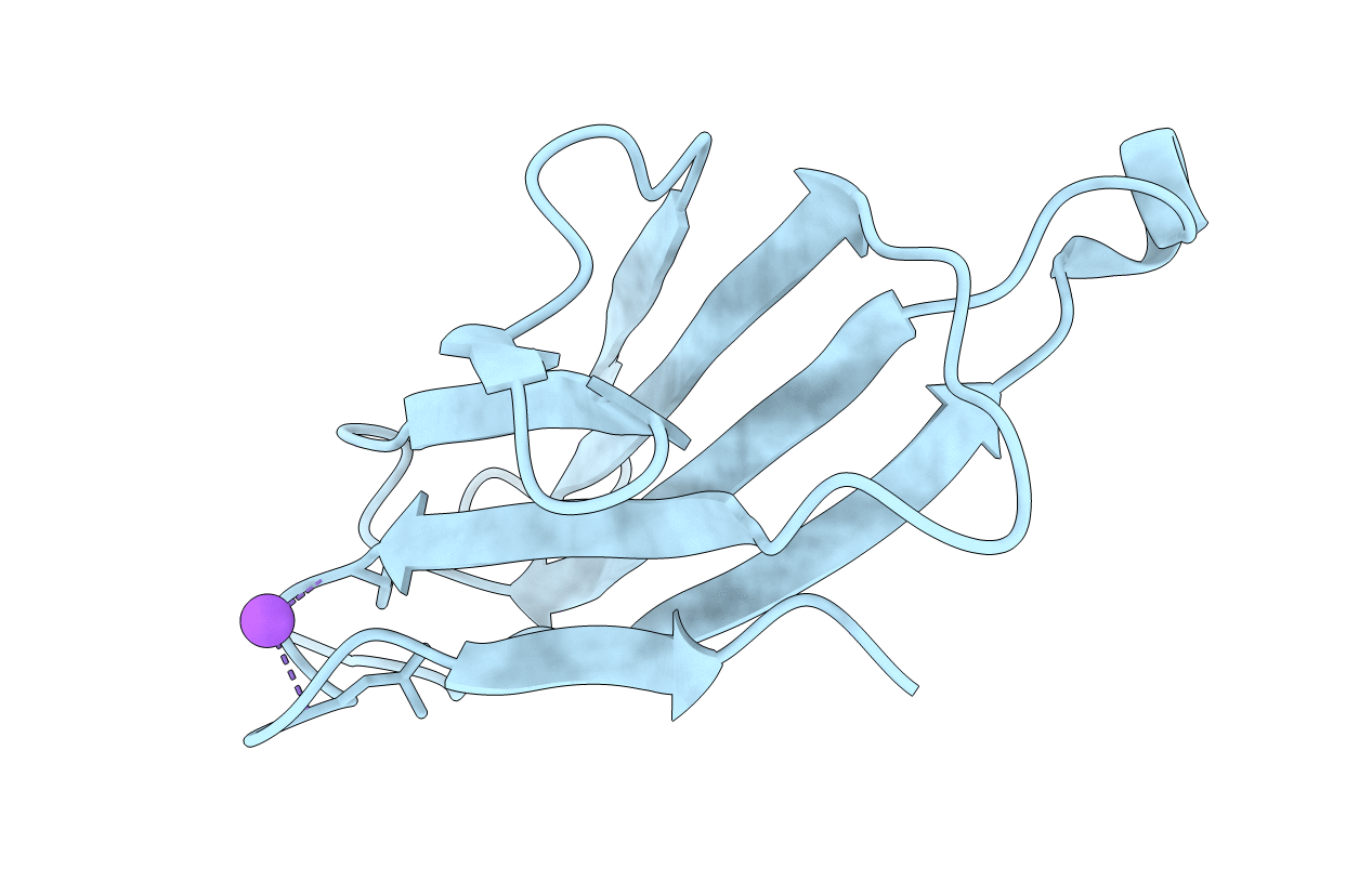
Crystal Structure of monomeric human beta-2-microglobulin
Biological Source:
Source Organism:
PDB Version:
Deposition Date:
2002-04-09
Release Date:
2002-07-31
Method Details:
Experimental Method:
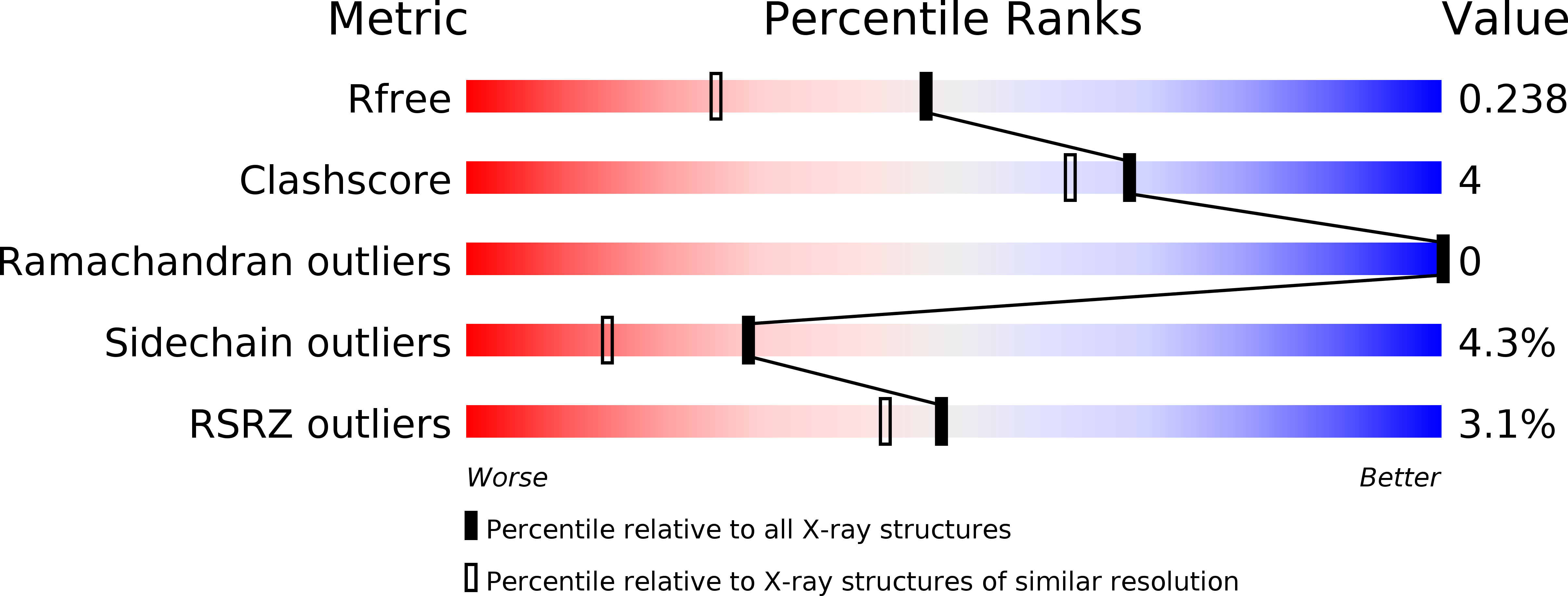
Resolution:
1.80 Å
R-Value Free:
0.23
R-Value Work:
0.18
Space Group:
C 1 2 1
Macromolecular Entities
Polymer Type:polypeptide(L)
Description:beta-2-microglobulin
Chain IDs:A
Chain Length:100
Number of Molecules:1
Biological Source:Homo sapiens
Ligand Molecules
Primary Citation
Crystal structure of monomeric human beta-2-microglobulin reveals clues to its amyloidogenic properties.
Proc.Natl.Acad.Sci.USA 99 9771 9776 (2002)
PMID: 12119416 DOI: 10.1073/pnas.152337399

Abstact

Dissociation of human beta-2-microglobulin (beta(2)m) from the heavy chain of the class I HLA complex is a critical first step in the formation of amyloid fibrils from this protein. As a consequence of renal failure, the concentration of circulating monomeric beta(2)m increases, ultimately leading to deposition of the protein into amyloid fibrils and development of the disorder, dialysis-related amyloidosis. Here we present the crystal structure of a monomeric form of human beta(2)m determined at 1.8-A resolution that reveals remarkable structural changes relative to the HLA-bound protein. These involve the restructuring of a beta bulge that separates two short beta strands to form a new six-residue beta strand at one edge of this beta sandwich protein. These structural changes remove key features proposed to have evolved to protect beta sheet proteins from aggregation [Richardson, J. & Richardson, D. (2002) Proc. Natl. Acad. Sci. USA 99, 2754-2759] and replaces them with an aggregation-competent surface. In combination with solution studies using (1)H NMR, we show that the crystal structure presented here represents a rare species in solution that could provide important clues about the mechanism of amyloid formation from the normally highly soluble native protein.

Legend

Protein

Chemical

Disease

Primary Citation of related structures