
Deposition Date
1995-03-20
Release Date
1996-03-08
Last Version Date
2024-10-30
Entry Detail
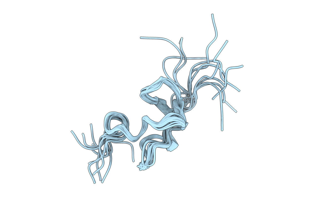
PDB ID:
1LDL
Keywords:
Title:
THREE-DIMENSIONAL STRUCTURE OF A CYSTEINE-RICH REPEAT FROM THE LOW-DENSITY LIPOPROTEIN RECEPTOR
Biological Source:
Source Organism(s):
Homo sapiens (Taxon ID: 9606)
Expression System(s):
Method Details:
Experimental Method:
Conformers Submitted:
10
