
Deposition Date
1994-01-11
Release Date
1994-08-31
Last Version Date
2024-11-13
Entry Detail
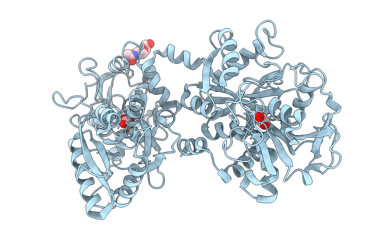
PDB ID:
1LCF
Keywords:
Title:
CRYSTAL STRUCTURE OF COPPER-AND OXALATE-SUBSTITUTED HUMAN LACTOFERRIN AT 2.0 ANGSTROMS RESOLUTION
Biological Source:
Source Organism(s):
Homo sapiens (Taxon ID: 9606)
Method Details:
Experimental Method:
Resolution:
2.00 Å
R-Value Observed:
0.19
Space Group:
P 21 21 21
