1LC3 image
Deposition Date 2002-04-05
Release Date 2002-07-17
Last Version Date 2024-02-14
Entry Detail
PDB ID:
1LC3
Keywords:
Title:
Crystal Structure of a Biliverdin Reductase Enzyme-Cofactor Complex
Biological Source:
Source Organism:
Host Organism:
Method Details:
Experimental Method:
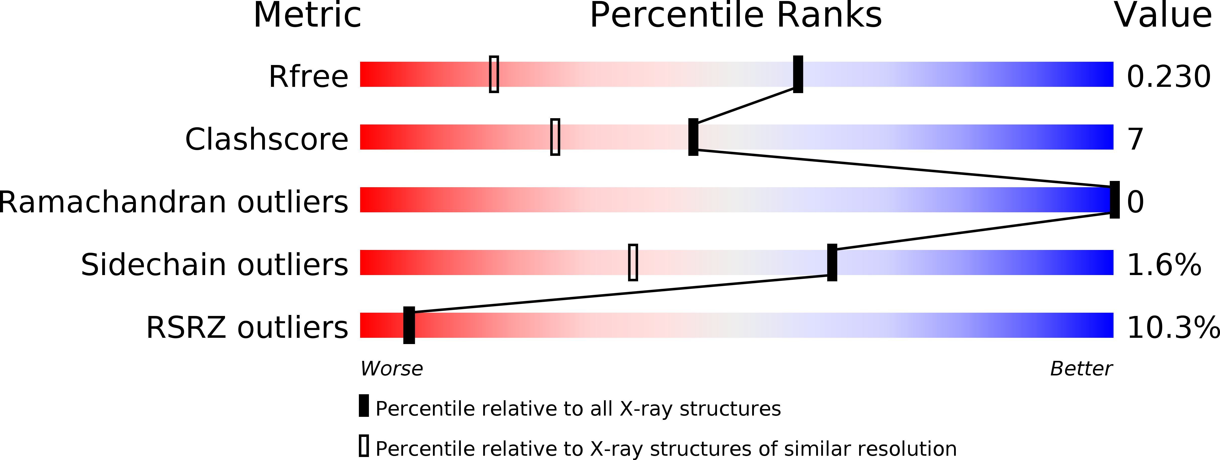
Resolution:
1.50 Å
R-Value Free:
0.23
R-Value Work:
0.21
Space Group:
P 21 21 21
Macromolecular Entities
Structures with similar UniProt ID
Protein Blast
Polymer Type:polypeptide(L)
Molecule:Biliverdin Reductase A
Gene (Uniprot):Blvra
Chain IDs:A
Chain Length:294
Number of Molecules:1
Biological Source:Rattus norvegicus
Primary Citation
Crystal structure of a biliverdin IXalpha reductase enzyme-cofactor complex.
J.Mol.Biol. 319 1199 1210 (2002)
PMID: 12079357 DOI: 10.1016/S0022-2836(02)00383-2

Abstact

Biliverdin reductase (BVR) catalyzes the last step in heme degradation by reducing the gamma-methene bridge of the open tetrapyrrole, biliverdin IXalpha, to bilirubin with the concomitant oxidation of a beta-nicotinamide adenine dinucleotide (NADH) or beta-nicotinamide adenine dinucleotide phosphate (NADPH) cofactor. Bilirubin is the major bile pigment in mammals and has antioxidant and anticompliment activity. We have determined X-ray crystal structures of apo rat BVR and its complex with NADH at 1.2 A and 1.5 A resolution, respectively. In agreement with an independent structure determination of the apo-enzyme, BVR consists of an N-terminal dinucleotide-binding domain (Rossmann-fold) and a C-terminal domain that contains a six-stranded beta-sheet that is flanked on one face by several alpha-helices. The C-terminal and N-terminal domains interact extensively, forming the active site cleft at their interface. The cofactor complex structure reported here reveals that the cofactor nicotinamide ring extends into the active site cleft, where it is adjacent to conserved amino acid residues and, consistent with the known stereochemistry of the reaction catalyzed by BVR, the si face of the ring is accessible for hydride transfer. The only titratable side-chain that appears to be suitably positioned to function as a general acid in catalysis is Tyr97. This residue, however, is not essential for catalysis, since the Tyr97Phe mutant protein retains 50% activity. This finding suggests that the dominant role in catalysis may be performed by hydride transfer from the cofactor, a process that may be promoted by proximity of the invariant residues Glu96, Glu123, and Glu126, to the nicotinamide ring.

Legend

Protein

Chemical

Disease

Primary Citation of related structures
Feedback Form
Name
Email
Institute
Feedback