1LBJ image
Deposition Date 2002-04-03
Release Date 2002-11-20
Last Version Date 2024-05-22
Entry Detail
PDB ID:
1LBJ
Title:
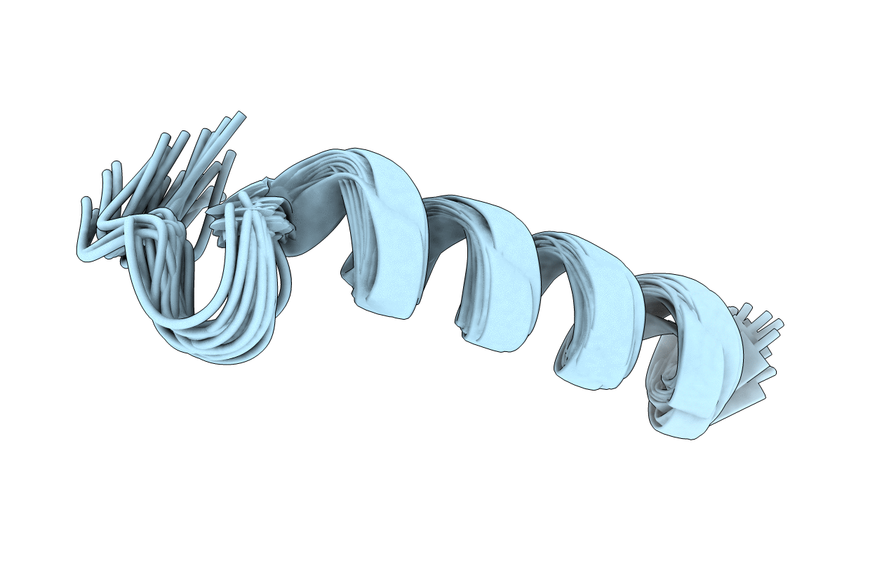
NMR solution structure of motilin in phospholipid bicellar solution
Biological Source:
Source Organism:
(Taxon ID: )
Method Details:
Experimental Method:
Conformers Calculated:
100
Conformers Submitted:
24
Selection Criteria:
structures with the least restraint violations,structures with the lowest energy
Macromolecular Entities
Polymer Type:polypeptide(L)
Molecule:motilin
Gene (Uniprot):MLN
Chain IDs:A
Chain Length:22
Number of Molecules:1
Biological Source:
Ligand Molecules
Primary Citation
NMR solution structure and dynamics of motilin in isotropic phospholipid bicellar solution
J.BIOMOL.NMR 24 103 112 (2002)
PMID: 12495026 DOI: 10.1023/A:1020902915969

Abstact

The structure and dynamics of the gastrointestinal peptide hormone motilin, consisting of 22 amino acid residues, have been studied in the presence of isotropic q = 0.5 phospholipid bicelles. The NMR solution structure of the peptide in acidic bicelle solution was determined from 203 NOE-derived distance constraints and six backbone torsion angle constraints. Dynamic properties for the 13Calpha-1H vector in Leu10 were determined for motilin specifically labeled with 13C at this position by analysis of multiple-field relaxation data. The structure reveals an ordered alpha-helical conformation between Glu9 and Lys20. The N-terminus is also well structured with a turn resembling that of a classical beta-turn. The 13C dynamics clearly show that motilin tumbles slowly in solution, with a correlation time characteristic of a large object. It was also found that motilin has a large degree of local flexibility as compared with what has previously been reported in SDS micelles. The results show that motilin interacts with the bicelle, displaying motional properties of a peptide bound to a membrane. In comparison, motilin in neutral bicelles seems less structured and more flexible. This study shows that the small isotropic bicelles are well suited for use as membrane-mimetic for structural as well as dynamical investigations of membrane-bound peptides by high-resolution NMR.

Legend

Protein

Chemical

Disease

Primary Citation of related structures