Deposition Date
2002-03-28
Release Date
2003-11-11
Last Version Date
2024-10-23
Entry Detail
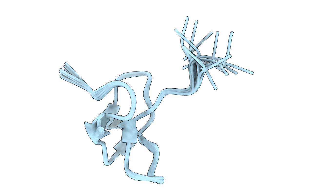
Method Details:
Experimental Method:
Conformers Calculated:
100
Conformers Submitted:
20
Selection Criteria:
structures with the least restraint violations,structures with the lowest energy