1LA3 image
Deposition Date 2002-03-27
Release Date 2002-06-19
Last Version Date 2024-10-30
Entry Detail
PDB ID:
1LA3
Title:
Solution structure of recoverin mutant, E85Q
Biological Source:
Source Organism:
Bos taurus (Taxon ID: 9913)
Host Organism:
Method Details:
Experimental Method:
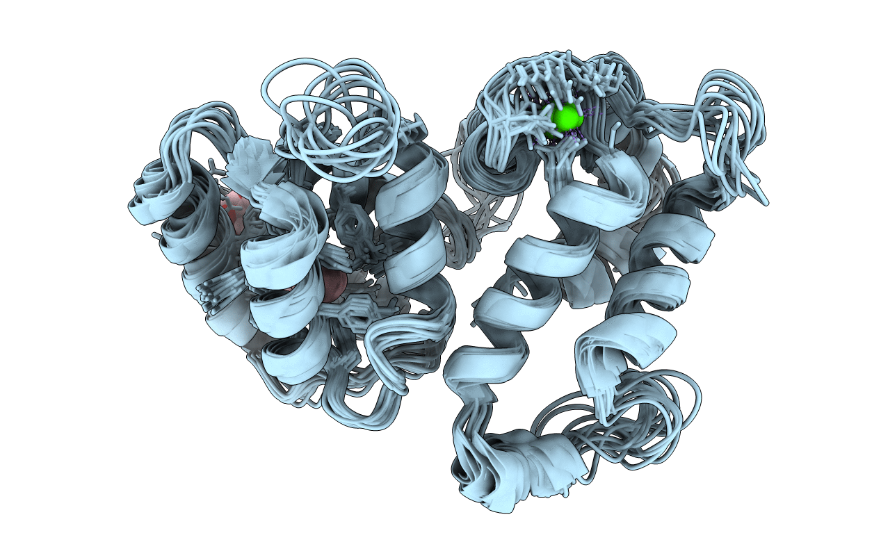
Conformers Calculated:
20
Conformers Submitted:
14
Selection Criteria:
The submitted conformer models are the 14 structures with lowest energy
Macromolecular Entities
Structures with similar UniProt ID
Protein Blast
Polymer Type:polypeptide(L)
Molecule:Recoverin
Gene (Uniprot):RCVRN
Mutagens:E85Q
Chain IDs:A
Chain Length:201
Number of Molecules:1
Biological Source:Bos taurus
Primary Citation
Structure and calcium-binding studies of a recoverin mutant (E85Q) in an allosteric intermediate state.
Biochemistry 41 5776 5787 (2002)
PMID: 11980481 DOI: 10.1021/bi012153k

Abstact

Recoverin, a member of the EF-hand superfamily, serves as a calcium sensor in retinal rod cells. A myristoyl or related fatty acyl group covalently attached to the N-terminus of recoverin facilitates the binding of recoverin to retinal disk membranes by a mechanism known as the Ca2+-myristoyl switch. Previous structural studies revealed that the myristoyl group of recoverin is sequestered inside the protein core in the absence of calcium. The cooperative binding of two calcium ions to the second and third EF-hands (EF-2 and EF-3) of recoverin leads to the extrusion of the fatty acid. Here we present nuclear magnetic resonance (NMR), fluorescence, and calcium-binding studies of a myristoylated recoverin mutant (myr-E85Q) designed to abolish high-affinity calcium binding to EF-2 and thereby trap the myristoylated protein with calcium bound solely to EF-3. Equilibrium calcium-binding studies confirm that only one Ca2+ binds to myr-E85Q under the conditions of this study with a dissociation constant of 100 microM. Fluorescence and NMR spectra of the Ca2+-free myr-E85Q are identical to those of Ca2+-free wild type, indicating that the E85Q mutation does not alter the stability and structure of the Ca2+-free protein. In contrast, the fluorescence and NMR spectra of half-saturated myr-E85Q (one bound Ca2+) look different from those of Ca2+-saturated wild type (two bound Ca2+), suggesting that half-saturated myr-E85Q may represent a structural intermediate. We report here the three-dimensional structure of Ca2+-bound myr-E85Q as determined by NMR spectroscopy. The N-terminal myristoyl group of Ca2+-bound myr-E85Q is sequestered within a hydrophobic cavity lined by many aromatic residues (F23, W31, Y53, F56, F83, and Y86) resembling that of Ca2+-free recoverin. The structure of Ca2+-bound myr-E85Q in the N-terminal region (residues 2-90) is similar to that of Ca2+-free recoverin, whereas the C-terminal region (residues 100-202) is more similar to that of Ca2+-bound wild type. Hence, the structure of Ca2+-bound myr-E85Q represents a hybrid between the structures of recoverin with zero and two Ca2+ bound. The binding of Ca2+ to EF-3 leads to local structural changes within the EF-hand that alter the domain interface and cause a 45 degrees swiveling of the N- and C-terminal domains, resulting in a partial unclamping of the myristoyl group. We propose that Ca2+-bound myr-E85Q may represent a stable intermediate state in the kinetic mechanism of the calcium-myristoyl switch.

Legend

Protein

Chemical

Disease

Primary Citation of related structures