Deposition Date
1992-02-11
Release Date
1993-10-31
Last Version Date
2024-02-14
Entry Detail
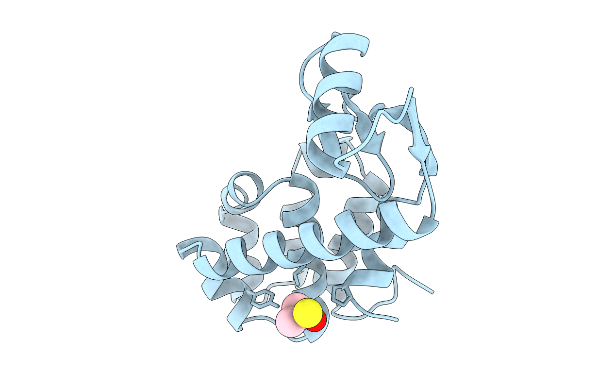
PDB ID:
1L96
Keywords:
Title:
STRUCTURE OF A HINGE-BENDING BACTERIOPHAGE T4 LYSOZYME MUTANT, ILE3-> PRO
Biological Source:
Source Organism(s):
Enterobacteria phage T4 (Taxon ID: 10665)
Method Details:
Experimental Method:
Resolution:
2.00 Å
R-Value Observed:
0.16
Space Group:
P 32 2 1