
Deposition Date
2002-03-21
Release Date
2002-06-26
Last Version Date
2024-10-30
Entry Detail
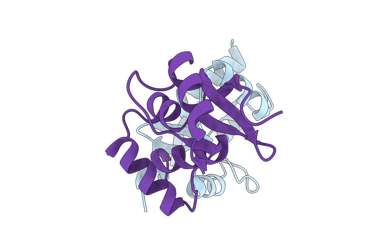
PDB ID:
1L8R
Keywords:
Title:
Structure of the Retinal Determination Protein Dachshund Reveals a DNA-Binding Motif
Biological Source:
Source Organism(s):
Homo sapiens (Taxon ID: 9606)
Expression System(s):
Method Details:
Experimental Method:
Resolution:
1.65 Å
R-Value Free:
0.25
R-Value Work:
0.23
R-Value Observed:
0.23
Space Group:
P 1
