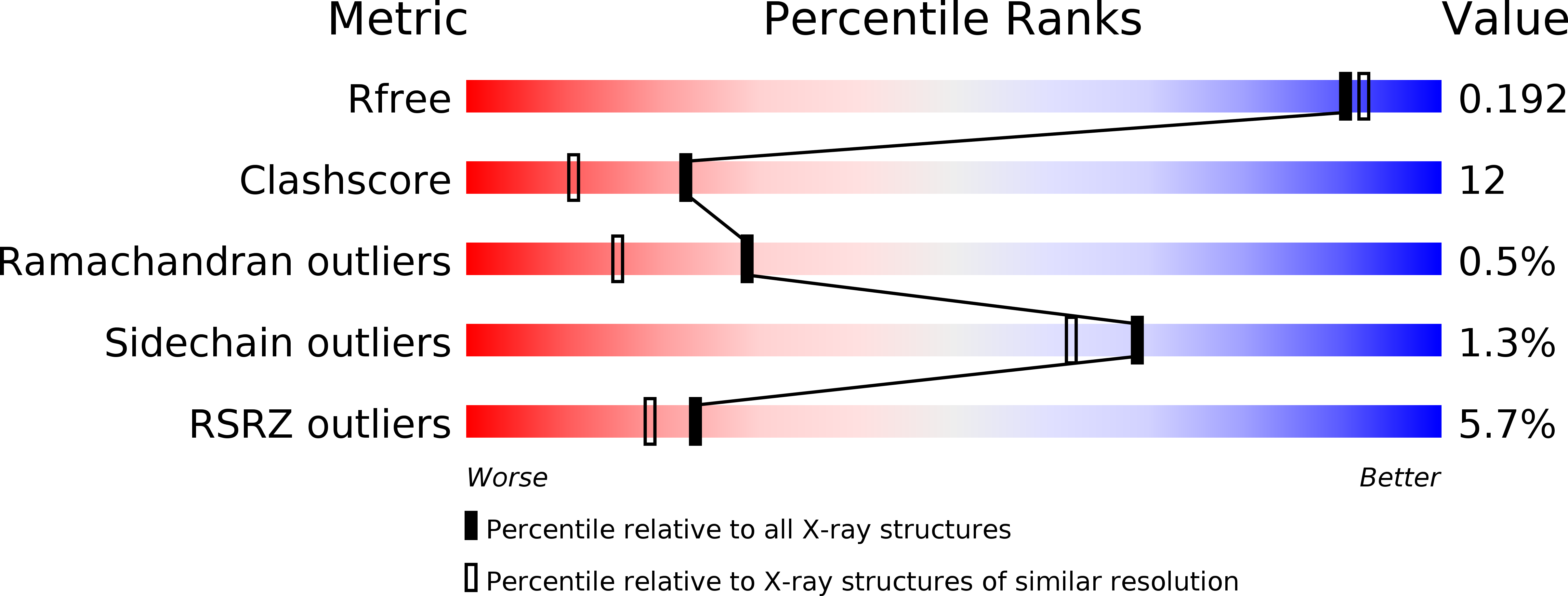
1L7I image
Deposition Date 2002-03-15
Release Date 2002-07-10
Last Version Date 2024-11-20
Entry Detail
PDB ID:
1L7I
Keywords:
Title:
Crystal Structure of the anti-ErbB2 Fab2C4
Biological Source:
Source Organism:
Homo sapiens (Taxon ID: 9606)
Host Organism:
Method Details:
Experimental Method:
Resolution:
1.80 Å
R-Value Free:
0.23
R-Value Work:
0.19
Space Group:
P 1 21 1
Macromolecular Entities
Polymer Type:polypeptide(L)
Molecule:chimera of Fab2C4: "humanized" murine monoclonal antibody
Chain IDs:B (auth: H)
Chain Length:222
Number of Molecules:1
Biological Source:Homo sapiens
Polymer Type:polypeptide(L)
Molecule:chimera of Fab2C4: "humanized" murine monoclonal antibody
Chain IDs:A (auth: L)
Chain Length:214
Number of Molecules:1
Biological Source:Homo sapiens
Ligand Molecules
Primary Citation
Comprehensive functional maps of the antigen-binding site of an anti-ErbB2 antibody obtained with shotgun scanning mutagenesis.
J.Mol.Biol. 320 415 428 (2002)
PMID: 12079396 DOI: 10.1016/S0022-2836(02)00264-4

Abstact

Shotgun scanning combinatorial mutagenesis was used to study the antigen-binding site of Fab2C4, a humanized monoclonal antibody fragment that binds to the extracellular domain of the human oncogene product ErbB2. Essentially all the residues in the Fab2C4 complementarity determining regions (CDRs) were alanine-scanned using phage-displayed libraries that preferentially allowed side-chains to vary as the wild-type or alanine. A separate homolog-scan was performed using libraries that allowed side-chains to vary only as the wild-type or a similar amino acid residue. Following binding selections to isolate functional clones, DNA sequencing was used to determine the wild-type/mutant ratios at each varied position, and these ratios were used to assess the contributions of each side-chain to antigen binding. The alanine-scan revealed that most of the side-chains that contribute to antigen binding are located in the heavy chain, and the Fab2C4 three-dimensional structure revealed that these residues fall into two groups. The first group consists of solvent-exposed residues which likely make energetically favorable contacts with the antigen and thus comprise the functional-binding epitope. The second group consists of buried residues with side-chains that pack against other CDR residues and apparently act as scaffolding to maintain the functional epitope in a binding-competent conformation. The homolog-scan involved subtle mutations, and as a result, only a subset of the side-chains that were intolerant to alanine substitutions were also intolerant to homologous substitutions. In particular, the 610 A2 functional epitope surface revealed by alanine-scanning shrunk to only 369 A2 when mapped with homologous substitutions, suggesting that this smaller subset of side-chains may be involved in more precise contacts with the antigen. The results validate shotgun scanning as a rapid and accurate method for determining the functional contributions of individual side-chains involved in protein-protein interactions.

Legend

Protein

Chemical

Disease

Primary Citation of related structures