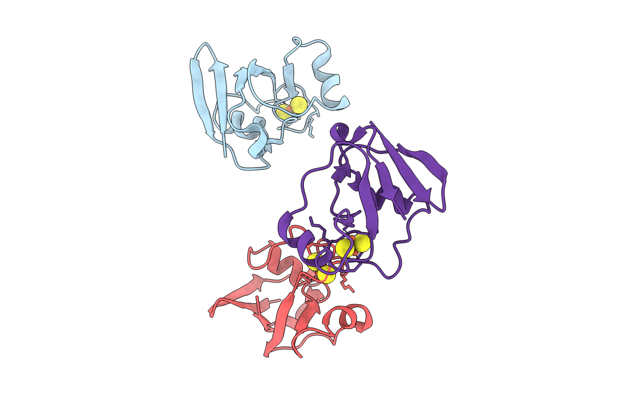
1L5P image
Deposition Date 2002-03-07
Release Date 2002-08-21
Last Version Date 2024-02-14
Entry Detail
PDB ID:
1L5P
Keywords:
Title:
Crystal Structure of Trichomonas vaginalis Ferredoxin
Biological Source:
Source Organism:
Method Details:
Experimental Method:
Resolution:
2.20 Å
R-Value Free:
0.31
R-Value Work:
0.24
Space Group:
P 21 21 21
Macromolecular Entities
Polymer Type:polypeptide(L)
Molecule:ferredoxin
Chain IDs:A, B, C
Chain Length:93
Number of Molecules:3
Biological Source:Trichomonas vaginalis
Ligand Molecules
Primary Citation
The crystal structure of Trichomonas vaginalis ferredoxin provides insight into metronidazole activation.
J.Mol.Biol. 318 503 518 (2002)
PMID: 12051855 DOI: 10.1016/S0022-2836(02)00051-7

Abstact

Crystallographic studies revealing the three-dimensional structure of the oxidized form of the [2Fe-2S] ferredoxin from Trichomonas vaginalis (TvFd) are presented. TvFd, a member of the hydrogenosomal class of ferredoxins, possesses a unique combination of redox and spectroscopic properties, and is believed to be the biological molecule that activates the drug metronidazole reductively in the treatment of trichomoniasis. It is the first hydrogenosomal ferredoxin to have its structure determined. The structure of TvFd reveals a monomeric, 93 residue protein with a fold similar to that of other known [2Fe-2S] ferredoxins. It contains nine hydrogen bonds to the sulfur atoms of the cluster, which is more than the number predicted on the basis of the spectroscopic data. The TvFd structure contains a large dipole moment like adrenodoxin, and appears to have a similar interaction domain. Our analysis demonstrates that TvFd has a unique cavity near the iron-sulfur cluster that exposes one of the inorganic sulfur atoms of the cluster to solvent. This cavity is not seen in any other [2Fe-2S] ferredoxin with known structure, and is hypothesized to be responsible for the high rate of metronidazole reduction by TvFd.

Legend

Protein

Chemical

Disease

Primary Citation of related structures