1L5B image
Deposition Date 2002-03-06
Release Date 2002-05-22
Last Version Date 2024-11-20
Entry Detail
PDB ID:
1L5B
Title:
DOMAIN-SWAPPED CYANOVIRIN-N DIMER
Biological Source:
Source Organism:
Host Organism:
Method Details:
Experimental Method:
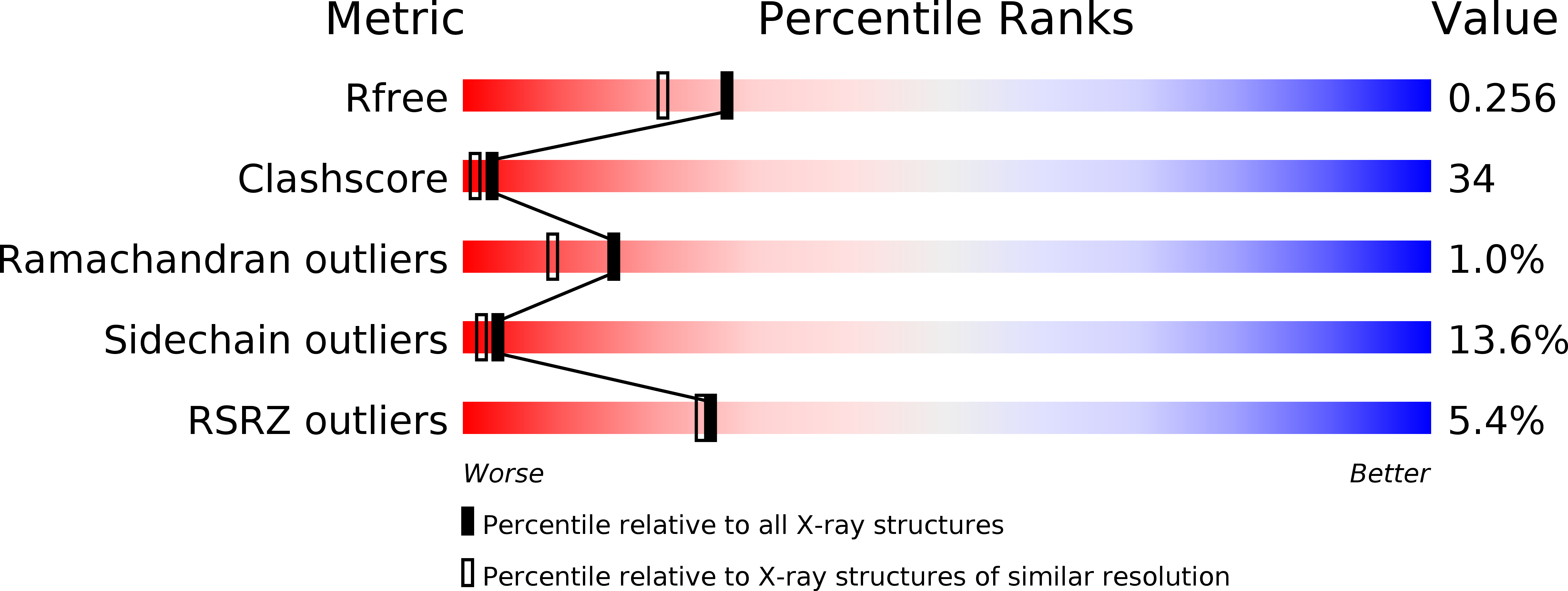
Resolution:
2.00 Å
R-Value Free:
0.25
R-Value Work:
0.24
Space Group:
P 41 21 2
Macromolecular Entities
Polymer Type:polypeptide(L)
Molecule:cyanovirin-N
Chain IDs:A, B
Chain Length:101
Number of Molecules:2
Biological Source:Nostoc ellipsosporum
Primary Citation
The domain-swapped dimer of cyanovirin-N is in a metastable folded state: reconciliation of X-ray and NMR structures.
Structure 10 673 686 (2002)
PMID: 12015150 DOI: 10.1016/S0969-2126(02)00758-X

Abstact

The structure of the potent HIV-inactivating protein cyanovirin-N was previously found by NMR to be a monomer in solution and a domain-swapped dimer by X-ray crystallography. Here we demonstrate that, in solution, CV-N can exist both in monomeric and in domain-swapped dimeric form. The dimer is a metastable, kinetically trapped structure at neutral pH and room temperature. Based on orientational NMR constraints, we show that the domain-swapped solution dimer is similar to structures in two different crystal forms, exhibiting solely a small reorientation around the hinge region. Mutation of the single proline residue in the hinge to glycine significantly stabilizes the protein in both its monomeric and dimeric forms. By contrast, mutation of the neighboring serine to proline results in an exclusively dimeric protein, caused by a drastic destabilization of the monomer.

Legend

Protein

Chemical

Disease

Primary Citation of related structures