Deposition Date
2002-03-06
Release Date
2002-06-12
Last Version Date
2023-08-30
Entry Detail
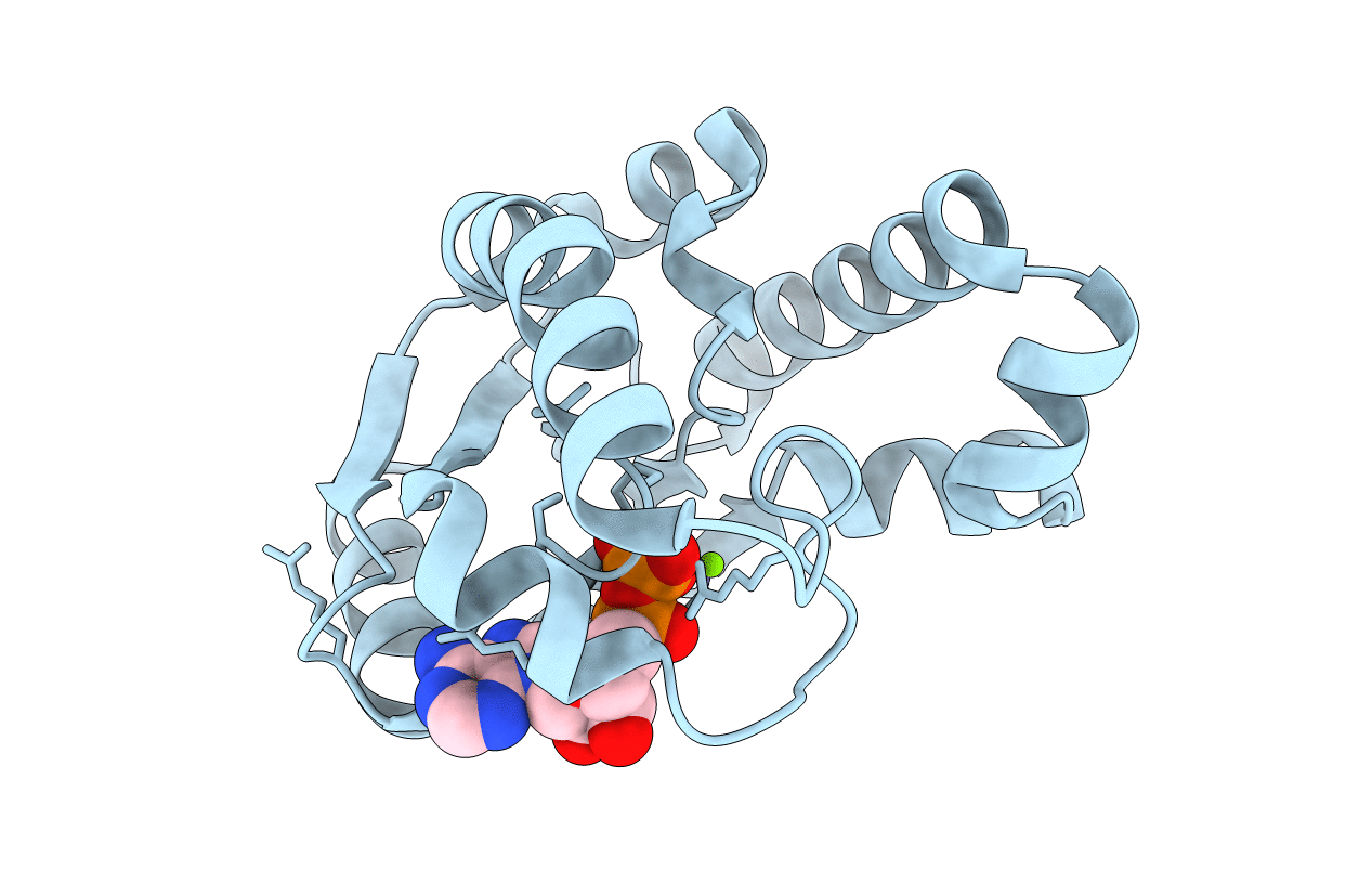
PDB ID:
1L4Y
Keywords:
Title:
CRYSTAL STRUCTURE OF SHIKIMATE KINASE FROM MYCOBACTERIUM TUBERCULOSIS IN COMPLEX WITH MGADP AT 2.0 ANGSTROM RESOLUTION
Biological Source:
Source Organism(s):
Mycobacterium tuberculosis (Taxon ID: 1773)
Expression System(s):
Method Details:
Experimental Method:
Resolution:
2.00 Å
R-Value Free:
0.25
R-Value Work:
0.21
Space Group:
P 32 2 1