Deposition Date
2002-03-04
Release Date
2002-07-31
Last Version Date
2023-08-16
Entry Detail
PDB ID:
1L4A
Keywords:
Title:
X-RAY STRUCTURE OF THE NEURONAL COMPLEXIN/SNARE COMPLEX FROM THE SQUID LOLIGO PEALEI
Biological Source:
Source Organism(s):
Loligo pealei (Taxon ID: 6621)
Expression System(s):
Method Details:
Experimental Method:
Resolution:
2.95 Å
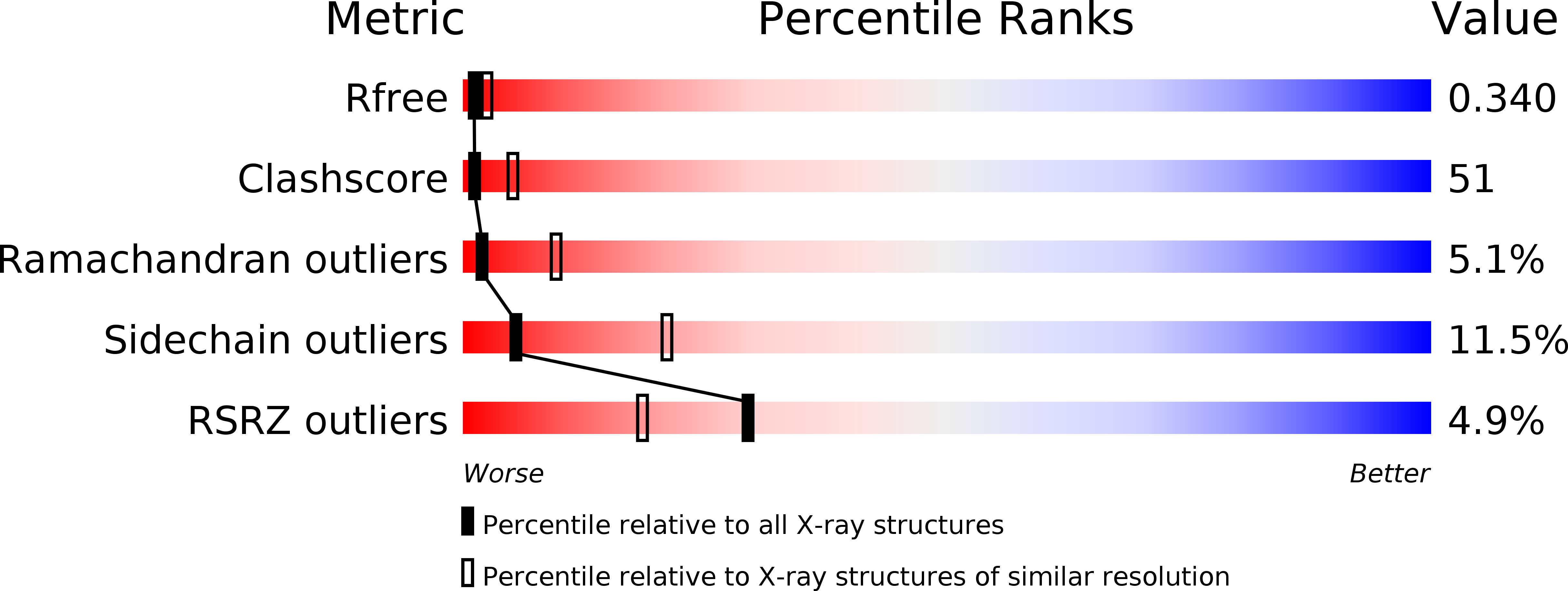
R-Value Free:
0.34
R-Value Work:
0.29
R-Value Observed:
0.30
Space Group:
P 21 21 21