Deposition Date
2002-03-01
Release Date
2002-04-26
Last Version Date
2024-12-25
Entry Detail
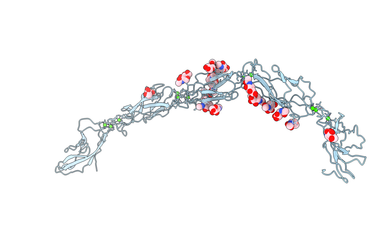
Biological Source:
Source Organism(s):
Xenopus laevis (Taxon ID: 8355)
Expression System(s):
Method Details:
Experimental Method:
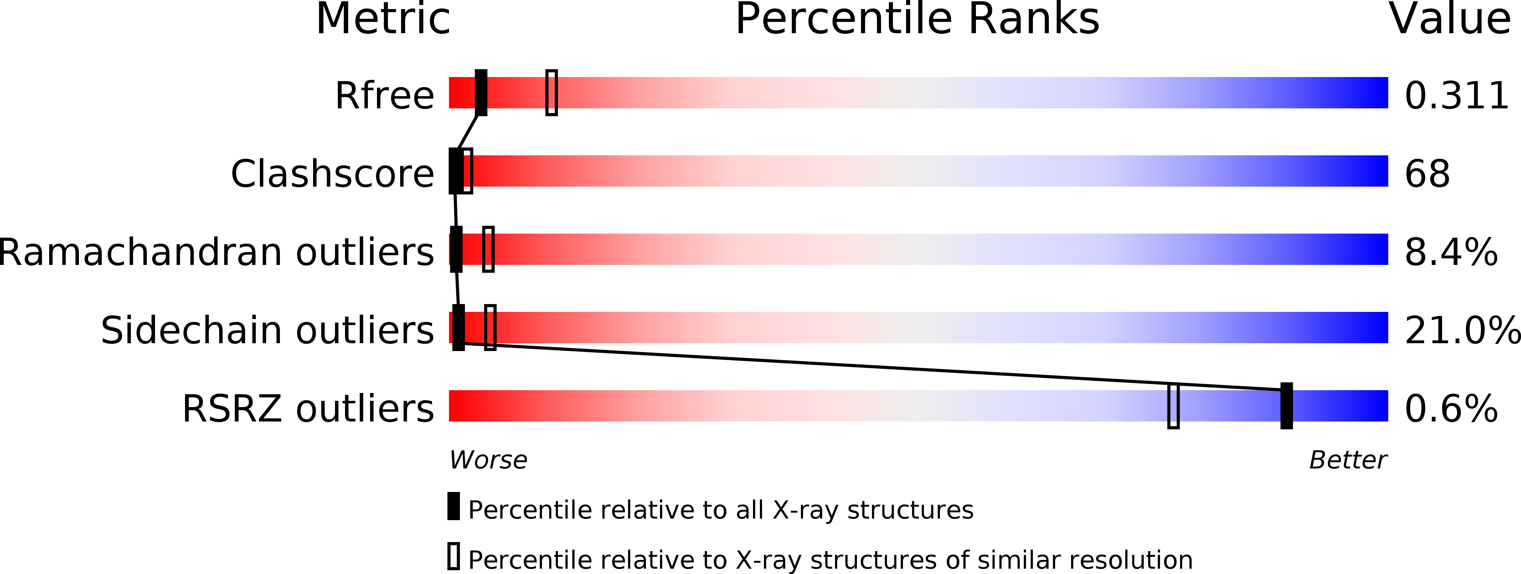
Resolution:
3.08 Å
R-Value Free:
0.27
R-Value Work:
0.24
R-Value Observed:
0.24
Space Group:
C 1 2 1