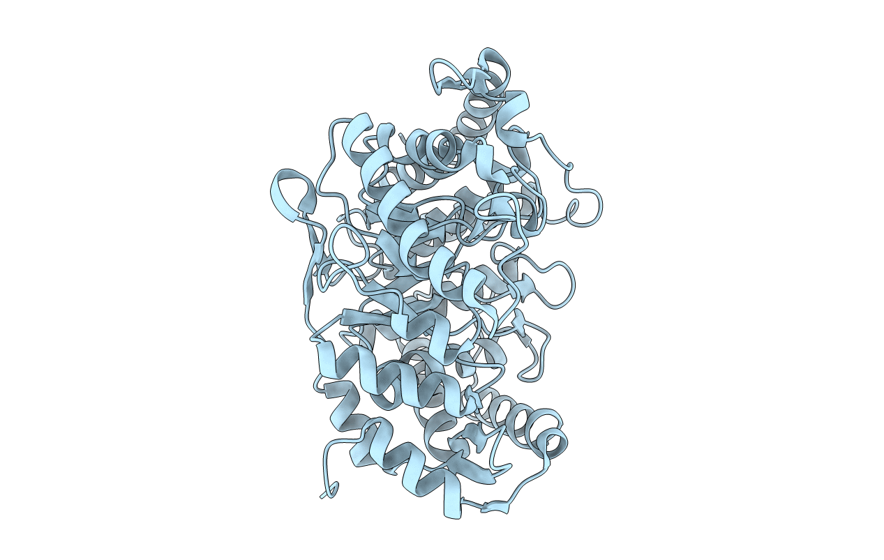
1L2Q image
Entry Detail
PDB ID:
1L2Q
Keywords:
Title:
Crystal Structure of the Methanosarcina barkeri Monomethylamine Methyltransferase (MtmB)
Biological Source:
Source Organism:
PDB Version:
Deposition Date:
2002-02-24
Release Date:
2002-06-05
Method Details:
Experimental Method:
Resolution:
1.70 Å
R-Value Free:
0.17
R-Value Work:
0.16
Space Group:
P 63 2 2
Macromolecular Entities
Polymer Type:polypeptide(L)
Description:monomethylamine methyltransferase
Chain IDs:A
Chain Length:458
Number of Molecules:1
Biological Source:Methanosarcina barkeri
Modified Residue
Compound ID Chain ID Parent Comp ID Details 2D Image
XPL A PYL ?
Ligand Molecules
Primary Citation
A new UAG-encoded residue in the structure of a methanogen methyltransferase.
Science 296 1462 1466 (2002)
PMID: 12029132 DOI: 10.1126/science.1069556

Abstact

Genes encoding methanogenic methylamine methyltransferases all contain an in-frame amber (UAG) codon that is read through during translation. We have identified the UAG-encoded residue in a 1.55 angstrom resolution structure of the Methanosarcina barkeri monomethylamine methyltransferase (MtmB). This structure reveals a homohexamer comprised of individual subunits with a TIM barrel fold. The electron density for the UAG-encoded residue is distinct from any of the 21 natural amino acids. Instead it appears consistent with a lysine in amide-linkage to (4R,5R)-4-substituted-pyrroline-5-carboxylate. We suggest that this amino acid be named l-pyrrolysine.

Legend

Protein

Chemical

Disease

Primary Citation of related structures