1L2F image
Deposition Date 2002-02-20
Release Date 2003-09-23
Last Version Date 2024-10-30
Entry Detail
PDB ID:
1L2F
Keywords:
Title:
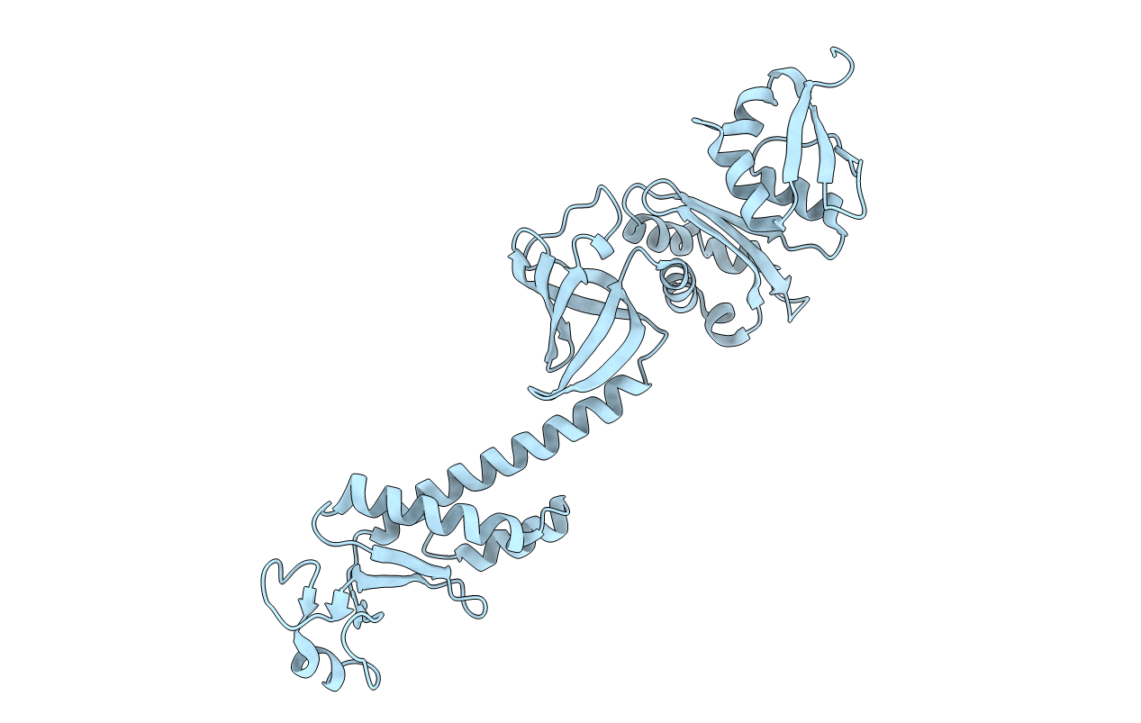
Crystal structure of NusA from Thermotoga maritima: a structure-based role of the N-terminal domain
Biological Source:
Source Organism:
Host Organism:
Method Details:
Experimental Method:
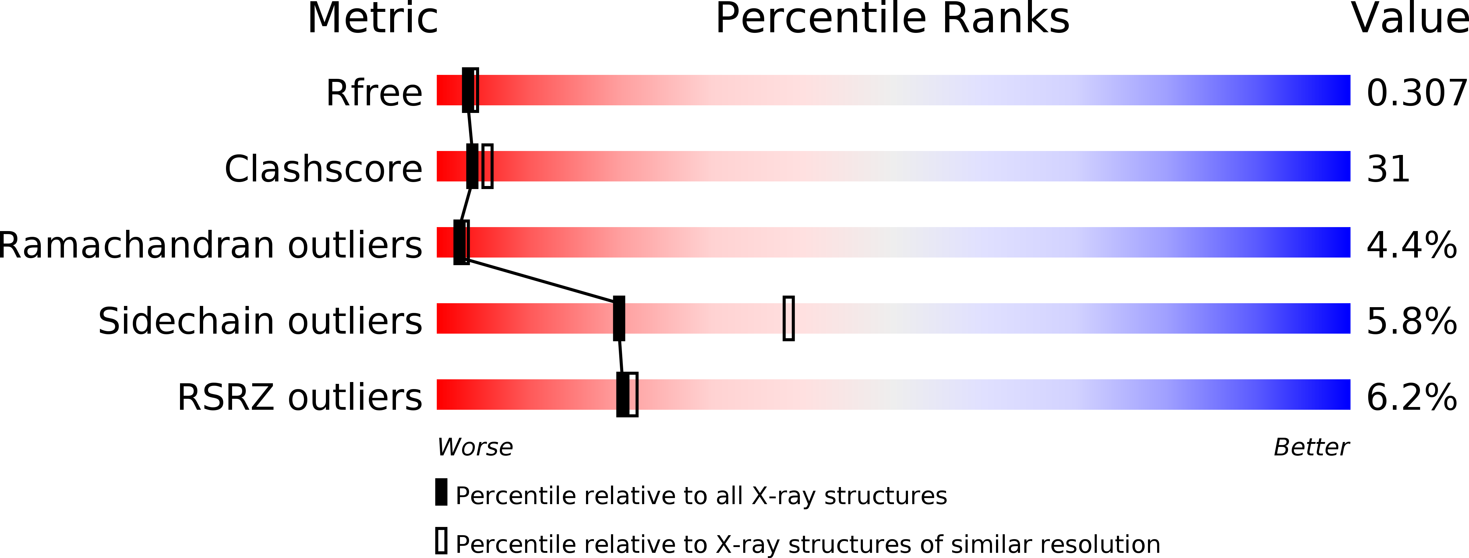
Resolution:
2.50 Å
R-Value Free:
0.29
R-Value Work:
0.22
Space Group:
P 43 21 2
Macromolecular Entities
Polymer Type:polypeptide(L)
Molecule:N utilization substance protein A
Gene (Uniprot):nusA
Chain IDs:A
Chain Length:369
Number of Molecules:1
Biological Source:Thermotoga maritima
Modified Residue
Compound ID Chain ID Parent Comp ID Details 2D Image
MSE A MET SELENOMETHIONINE
Primary Citation
Crystal structure of NusA from Thermotoga maritima and functional implication of the N-terminal domain.
Biochemistry 42 13429 13437 (2003)
PMID: 14621988 DOI: 10.1021/bi035118h

Abstact

We report the crystal structure of N-utilizing substance A protein (NusA) from Thermotoga maritima (TmNusA), a protein involved in transcriptional pausing, termination, and antitermination. TmNusA has an elongated rod-shaped structure consisting of an N-terminal domain (NTD, residues 1-132) and three RNA binding domains (RBD). The NTD consists of two subdomains, the globular head and the helical body domains, that comprise a unique three-dimensional structure that may be important for interacting with RNA polymerase. The globular head domain possesses a high content of negatively charged residues that may interact with the positively charged flaplike domain of RNA polymerase. The helical body domain is composed of a three-helix bundle that forms a hydrophobic core with the aid of two neighboring beta-strands. This domain shows structural similarity with one of the helical domains of sigma(70) factor from Escherichia coli. One side of the molecular surface shows positive electrostatic potential suitable for nonspecific RNA interaction. The RBD is composed of one S1 domain and two K-homology (KH) domains forming an elongated RNA binding surface. Structural comparison between TmNusA and Mycobacterium tuberculosis NusA reveals a possible hinge motion between NTD and RBD. In addition, a functional implication of the NTD in its interaction with RNA polymerase is discussed.

Legend

Protein

Chemical

Disease

Primary Citation of related structures