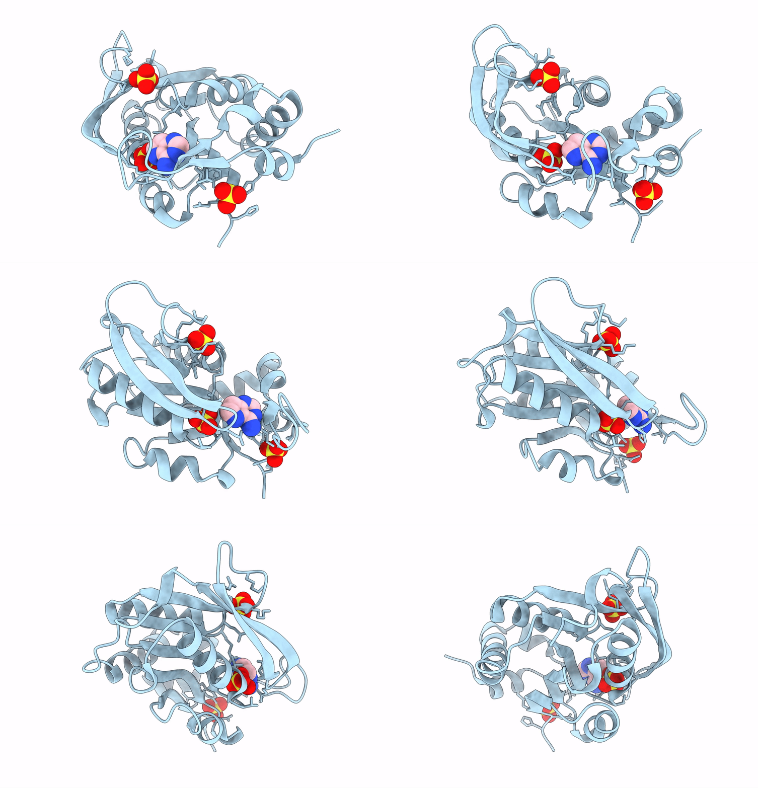
1L1Q image
Deposition Date 2002-02-19
Release Date 2002-11-27
Last Version Date 2023-08-16
Entry Detail
PDB ID:
1L1Q
Keywords:
Title:
Crystal Structure of APRTase from Giardia lamblia Complexed with 9-deazaadenine
Biological Source:
Source Organism:
Host Organism:
Method Details:
Experimental Method:
Resolution:
1.85 Å
R-Value Free:
0.25
R-Value Work:
0.21
R-Value Observed:
0.22
Space Group:
P 31 2 1
Macromolecular Entities
Polymer Type:polypeptide(L)
Molecule:Adenine phosphoribosyltransferase
Gene (Uniprot):DHA2_112885
Chain IDs:A
Chain Length:186
Number of Molecules:1
Biological Source:Giardia intestinalis
Primary Citation
Closed Site Complexes of Adenine Phosphoribosyltransferase from Giardia lamblia Reveal a Mechanism of Ribosyl Migration.
J.Biol.Chem. 277 39981 39988 (2002)
PMID: 12171925 DOI: 10.1074/jbc.M205596200

Abstact

The adenine phosphoribosyltransferase (APRTase) from Giardia lamblia was co-crystallized with 9-deazaadenine and sulfate or with 9-deazaadenine and Mg-phosphoribosylpyrophosphate. The complexes were solved and refined to 1.85 and 1.95 A resolution. Giardia APRTase is a symmetric homodimer with the monomers built around Rossman fold cores, an element common to all known purine phosphoribosyltransferases. The catalytic sites are capped with a small hood domain that is unique to the APRTases. These structures reveal several features relevant to the catalytic function of APRTase: 1) a non-proline cis peptide bond (Glu(61)-Ser(62)) is required to form the pyrophosphate binding site in the APRTase.9dA.MgPRPP complex but is a trans peptide bond in the absence of pyrophosphate group, as observed in the APRTase.9dA.SO4 complex; 2) a catalytic site loop is closed and fully ordered in both complexes, with Glu(100) from the catalytic loop acting as the acid/base for protonation/deprotonation of N-7 of the adenine ring; 3) the pyrophosphoryl charge is neutralized by a single Mg2+ ion and Arg(63), in contrast to the hypoxanthine-guanine phosphoribosyltransferases, which use two Mg2+ ions; and 4) the nearest structural neighbors to APRTases are the orotate phosphoribosyltransferases, suggesting different paths of evolution for adenine relative to other purine PRTases. An overlap comparison of AMP and 9-deazaadenine plus Mg-PRPP at the catalytic sites of APRTases indicated that reaction coordinate motion involves a 2.1-A excursion of the ribosyl anomeric carbon, whereas the adenine ring and the 5-phosphoryl group remained fixed. G. lamblia APRTase therefore provides another example of nucleophilic displacement by electrophile migration.

Legend

Protein

Chemical

Disease

Primary Citation of related structures