Deposition Date
2002-02-19
Release Date
2002-06-05
Last Version Date
2024-02-14
Entry Detail
PDB ID:
1L1O
Keywords:
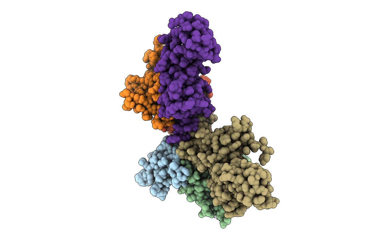
Title:
Structure of the human Replication Protein A (RPA) trimerization core
Biological Source:
Source Organism:
Homo sapiens (Taxon ID: 9606)
Host Organism:
Method Details:
Experimental Method:
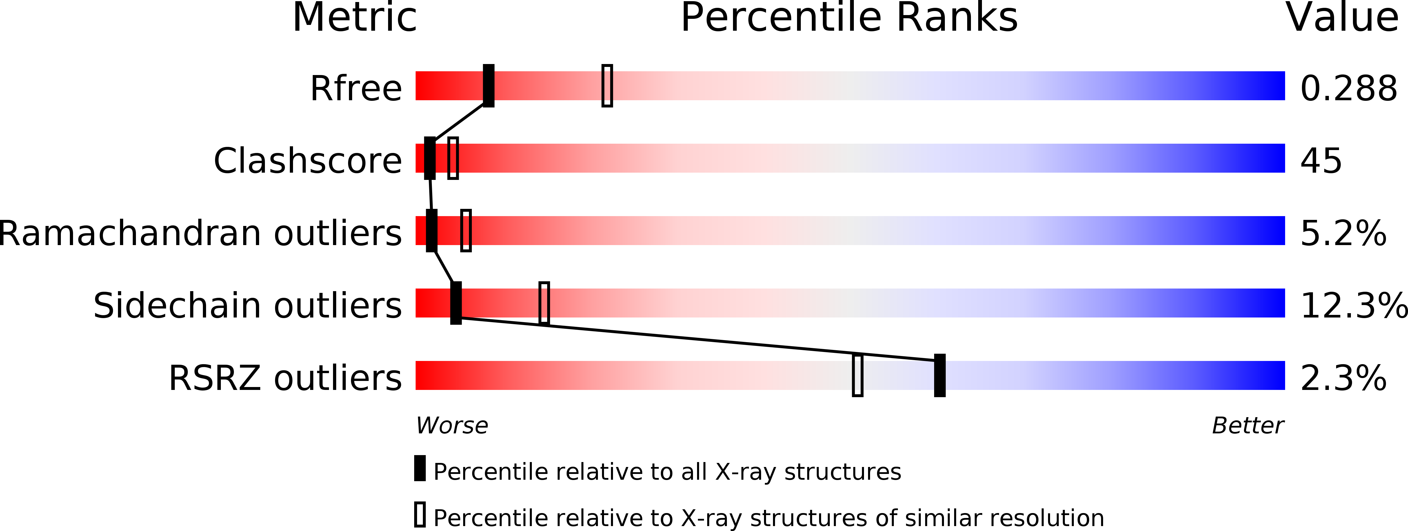
Resolution:
2.80 Å
R-Value Free:
0.28
R-Value Work:
0.23
R-Value Observed:
0.23
Space Group:
P 31 2 1