Deposition Date
2002-02-05
Release Date
2002-04-10
Last Version Date
2024-11-20
Entry Detail
PDB ID:
1KYR
Keywords:
Title:
Crystal Structure of a Cu-bound Green Fluorescent Protein Zn Biosensor
Biological Source:
Source Organism(s):
Aequorea victoria (Taxon ID: 6100)
Expression System(s):
Method Details:
Experimental Method:
Resolution:
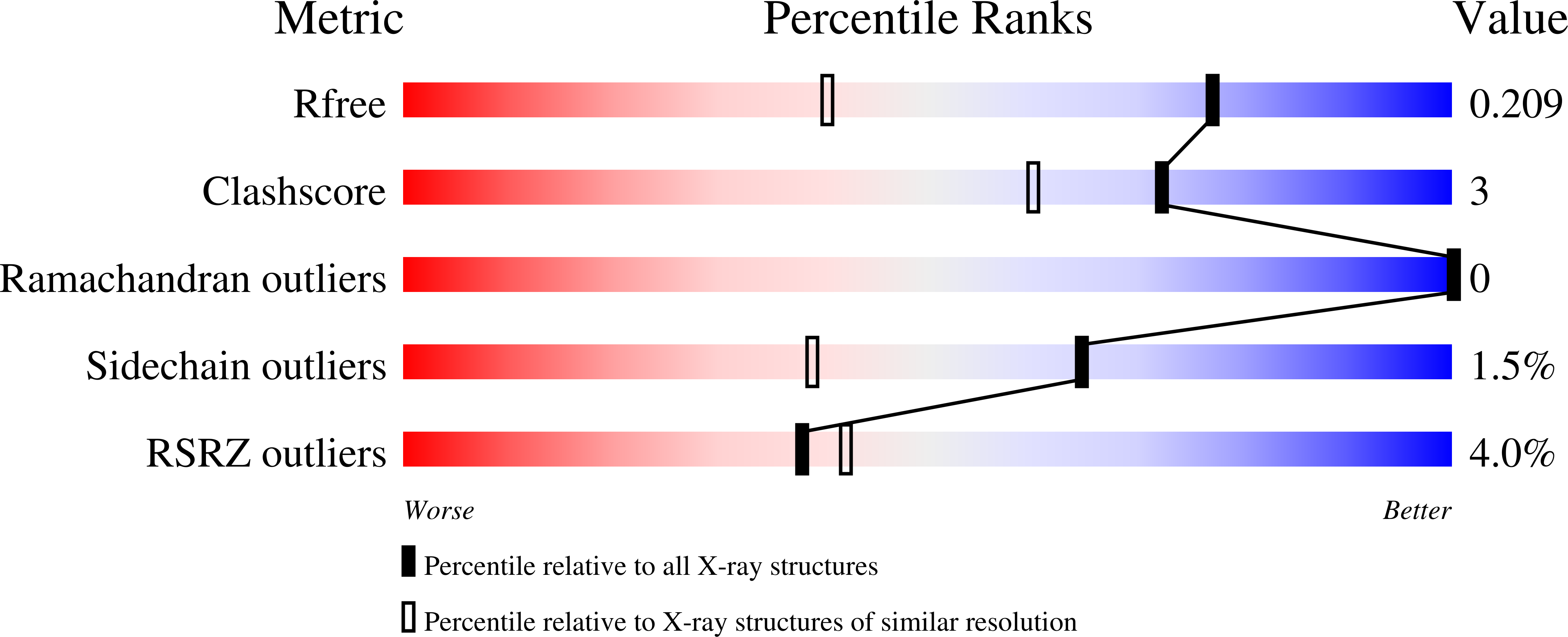
1.50 Å
R-Value Free:
0.21
R-Value Work:
0.15
R-Value Observed:
0.15
Space Group:
P 21 21 21