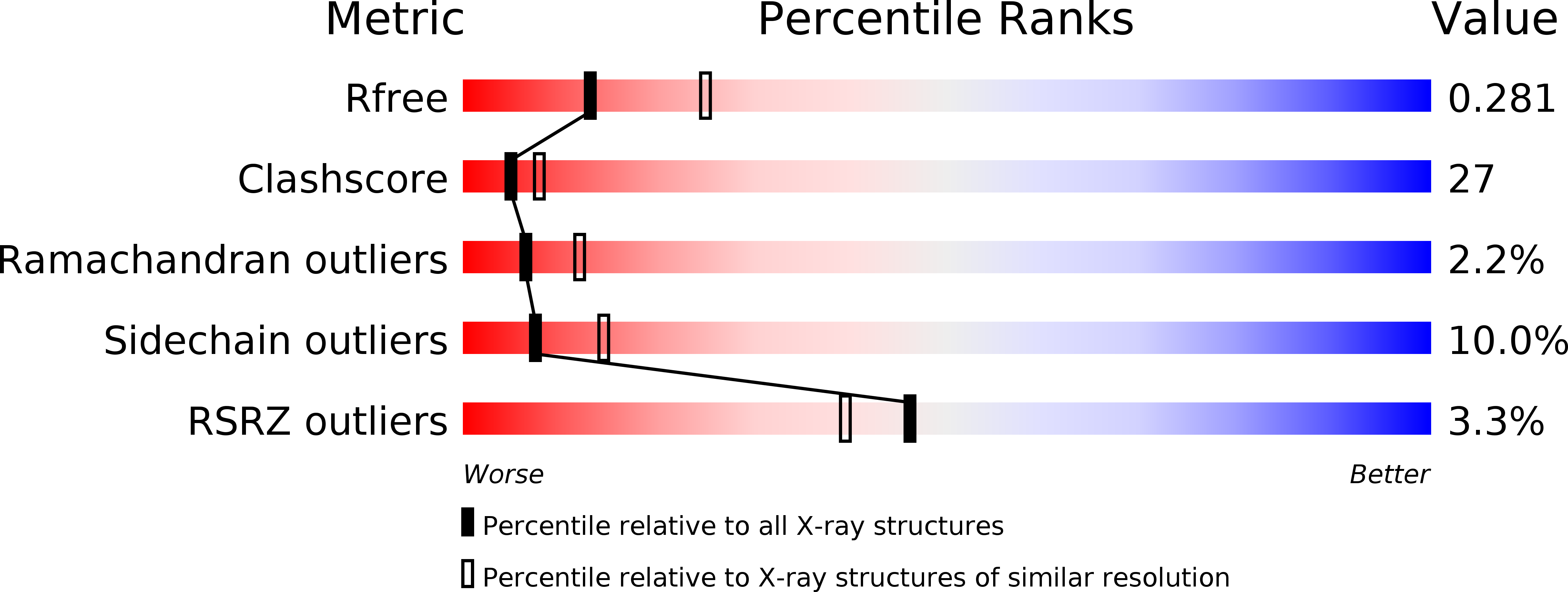
1KXU image
Deposition Date 1996-08-08
Release Date 1997-01-27
Last Version Date 2024-02-14
Entry Detail
PDB ID:
1KXU
Title:
CYCLIN H, A POSITIVE REGULATORY SUBUNIT OF CDK ACTIVATING KINASE
Biological Source:
Source Organism:
Homo sapiens (Taxon ID: 9606)
Host Organism:
Method Details:
Experimental Method:
Resolution:
2.60 Å
R-Value Free:
0.29
R-Value Work:
0.21
R-Value Observed:
0.21
Space Group:
I 41 2 2
Macromolecular Entities
Structures with similar UniProt ID
Protein Blast
Polymer Type:polypeptide(L)
Molecule:CYCLIN H
Gene (Uniprot):CCNH
Mutagens:INS(MSHHHHHHGS-M1) (HISTIDINE TAG)
Chain IDs:A
Chain Length:333
Number of Molecules:1
Biological Source:Homo sapiens
Primary Citation
Three-dimensional structure of human cyclin H, a positive regulator of the CDK-activating kinase.
Nat.Struct.Biol. 3 849 855 (1996)
PMID: 8836101 DOI: 10.1038/nsb1096-849

Abstact

Cyclin-dependent kinases (CDKs), which play a key role in cell cycle control, are activated by the CDK activating kinase (CAK), which activates cyclin-bound CDKs by phosphorylation at a specific threonine residue. Vertebrate CAK contains two key components: a kinase subunit with homology to its substrate CDKs and a regulatory subunit with homology to cyclins. We have determined the X-ray crystal structure of the regulatory subunit of CAK, cyclin H, at 2.6 A resolution. Cyclin H contains two alpha-helical core domains with a fold similar to that of cyclin A, a regulatory subunit of CAK substrate CDK2, and of TFIIB, a transcription factor. Outside of the core domains, the N- and C-terminal regions of the three structures are completely different. The conformational differences between cyclin H and A structures may reflect functional differences between the two cyclins.

Legend

Protein

Chemical

Disease

Primary Citation of related structures