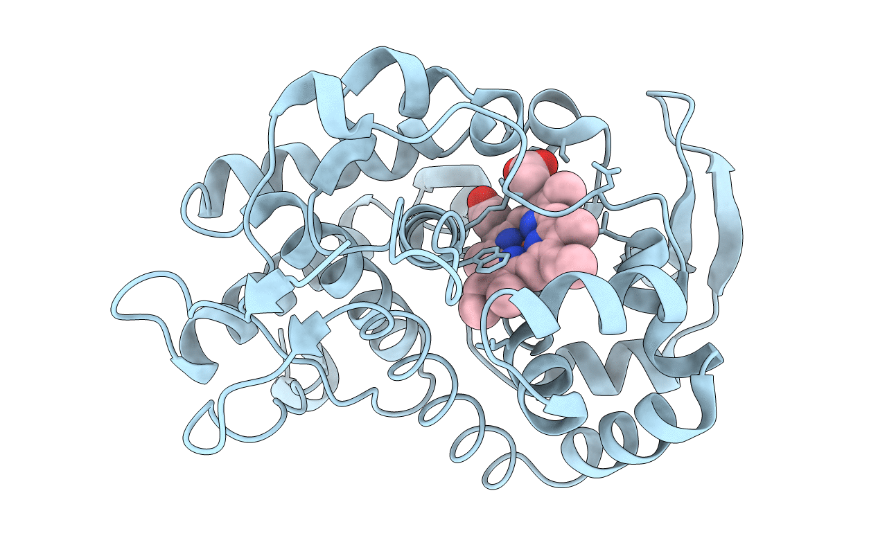
1KXN image
Entry Detail
PDB ID:
1KXN
Keywords:
Title:
Crystal Structure of Cytochrome c Peroxidase with a Proposed Electron Transfer Pathway Excised to Form a Ligand Binding Channel.
Biological Source:
Source Organism:
PDB Version:
Deposition Date:
2002-02-01
Release Date:
2002-03-06
Method Details:
Experimental Method:
Resolution:
1.80 Å
R-Value Free:
0.19
R-Value Work:
0.18
Space Group:
P 21 21 21
Macromolecular Entities
Polymer Type:polypeptide(L)
Description:cytochrome c peroxidase
Mutations:P190G
Chain IDs:A
Chain Length:289
Number of Molecules:1
Biological Source:Saccharomyces cerevisiae
Ligand Molecules
Primary Citation
Excision of a proposed electron transfer pathway in cytochrome c peroxidase and its replacement by a ligand-binding channel.
Protein Sci. 11 1251 1259 (2002)
PMID: 11967381 DOI: 10.1110/ps.4870102

Abstact

A previously proposed electron transfer (ET) pathway in the heme enzyme cytochrome c peroxidase has been excised from the structure, leaving an open ligand-binding channel in its place. Earlier studies on cavity mutants of this enzyme have revealed structural plasticity in this region of the molecule. Analysis of these structures has allowed the design of a variant in which the specific section of protein backbone representing a previously proposed ET pathway is accurately extracted from the protein. A crystal structure verified the creation of an open channel that overlays the removed segment, extending from the surface of the protein to the heme at the core of the protein. A number of heterocyclic cations were found to bind to the proximal-channel mutant with affinities that can be rationalized based on the structures. It is proposed that small ligands bind more weakly to the proximal-channel mutant than to the W191G cavity due to an increased off rate of the open channel, whereas larger ligands are able to bind to the channel mutant without inducing large conformational changes. The structure of benzimidazole bound to the proximal-channel mutant shows that the ligand accurately overlays the position of the tryptophan radical center that was removed from the wild-type enzyme and displaces four of the eight ordered solvent molecules seen in the empty cavity. Ligand binding also caused a small rearrangement of the redesigned protein loop, perhaps as a result of improved electrostatic interactions with the ligand. The engineered channel offers the potential for introducing synthetic replacements for the removed structure, such as sensitizer-linked substrates. These installed "molecular wires" could be used to rapidly initiate reactions, trap reactive intermediates, or answer unresolved questions about ET pathways.

Legend

Protein

Chemical

Disease

Primary Citation of related structures