1KWK image
Deposition Date 2002-01-29
Release Date 2002-10-02
Last Version Date 2024-03-13
Entry Detail
PDB ID:
1KWK
Keywords:
Title:
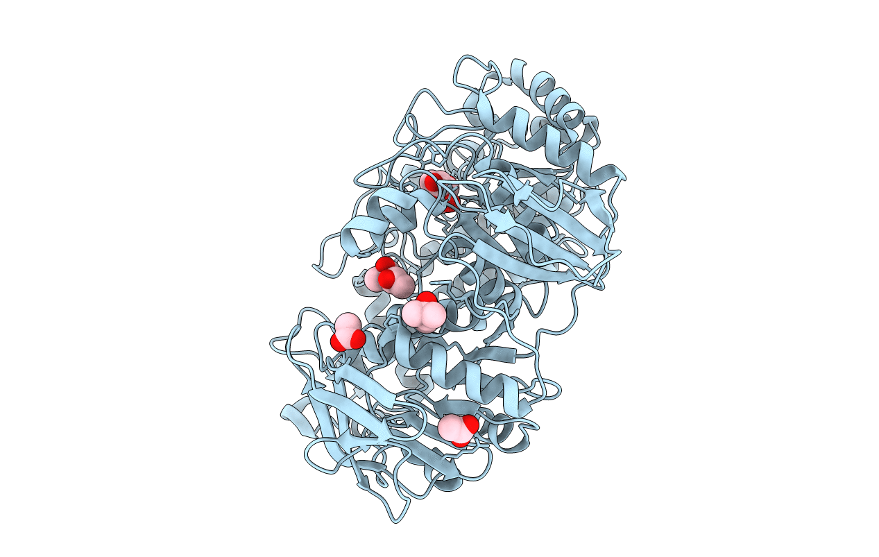
Crystal structure of Thermus thermophilus A4 beta-galactosidase in complex with galactose
Biological Source:
Source Organism:
Host Organism:
Method Details:
Experimental Method:
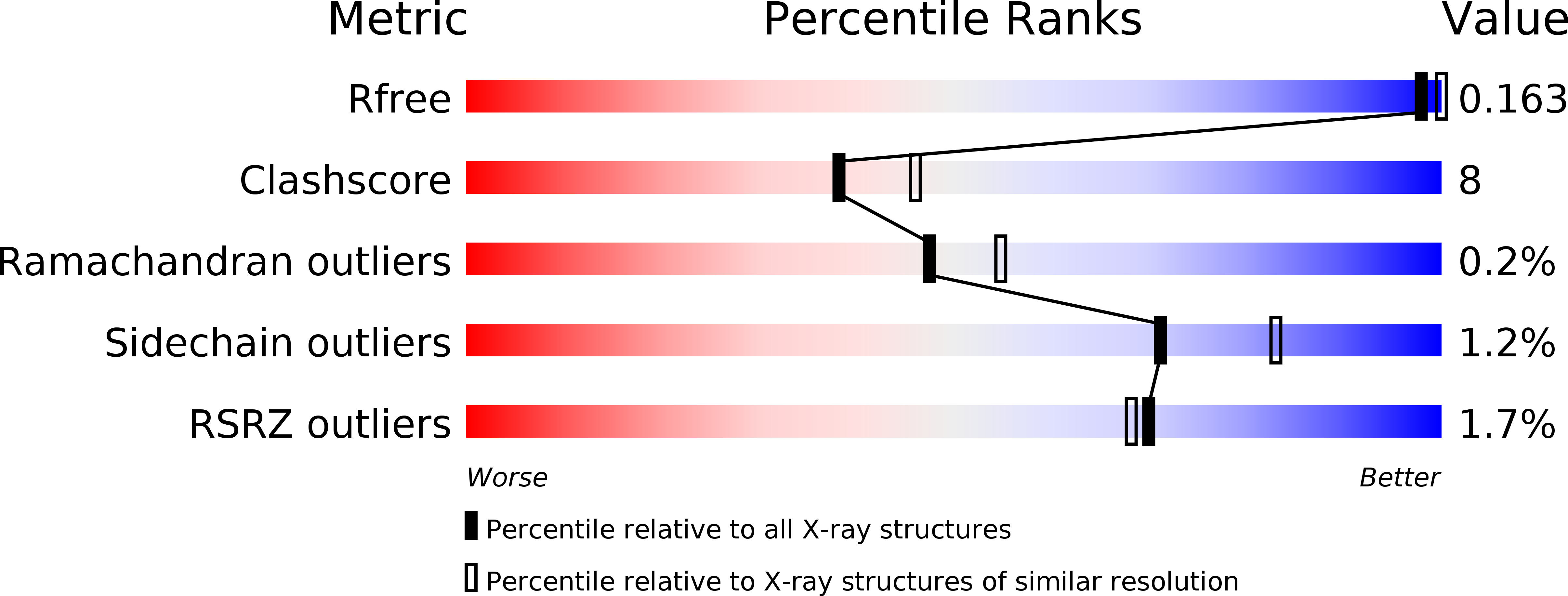
Resolution:
2.20 Å
R-Value Free:
0.20
R-Value Work:
0.16
R-Value Observed:
0.16
Space Group:
P 3 2 1
Macromolecular Entities
Polymer Type:polypeptide(L)
Molecule:BETA-GALACTOSIDASE
Chain IDs:A
Chain Length:645
Number of Molecules:1
Biological Source:Thermus thermophilus
Primary Citation
Trimeric crystal structure of the glycoside hydrolase family 42 beta-galactosidase from Thermus thermophilus A4 and the structure of its complex with galactose.
J.Mol.Biol. 322 79 91 (2002)
PMID: 12215416 DOI: 10.1016/S0022-2836(02)00746-5

Abstact

The beta-galactosidase from an extreme thermophile, Thermus thermophilus A4 (A4-beta-Gal), is thermostable and belongs to the glycoside hydrolase family 42 (GH-42). As the first known structures of a GH-42 enzyme, we determined the crystal structures of free and galactose-bound A4-beta-Gal at 1.6A and 2.2A resolution, respectively. A4-beta-Gal forms a homotrimeric structure resembling a flowerpot. Each monomer has an active site located inside a large central tunnel. The N-terminal domain of A4-beta-Gal has a TIM barrel fold, as predicted from hydrophobic cluster analysis. The putative catalytic residues of A4-beta-Gal (Glu141 and Glu312) superimpose well with the catalytic residues of Escherichia coli beta-galactosidase. The environment around the catalytic nucleophile (Glu312) is similar to that in the case of E.coli beta-galactosidase, but the recognition mechanism for a substrate is different. Trp182 of the next subunit of the trimer constitutes a part of the active-site pocket, indicating that the trimeric structure is essential for the enzyme activity. Structural comparison with other glycoside hydrolases revealed that many features of the 4/7 superfamily are conserved in the A4-beta-Gal structure. On the basis of the results of 1H NMR spectroscopy, A4-beta-Gal was determined to be a "retaining" enzyme. Interestingly, the active site was similar with those of retaining enzymes, but the overall fold of the TIM barrel domain was very similar to that of an inverting enzyme, beta-amylase.

Legend

Protein

Chemical

Disease

Primary Citation of related structures