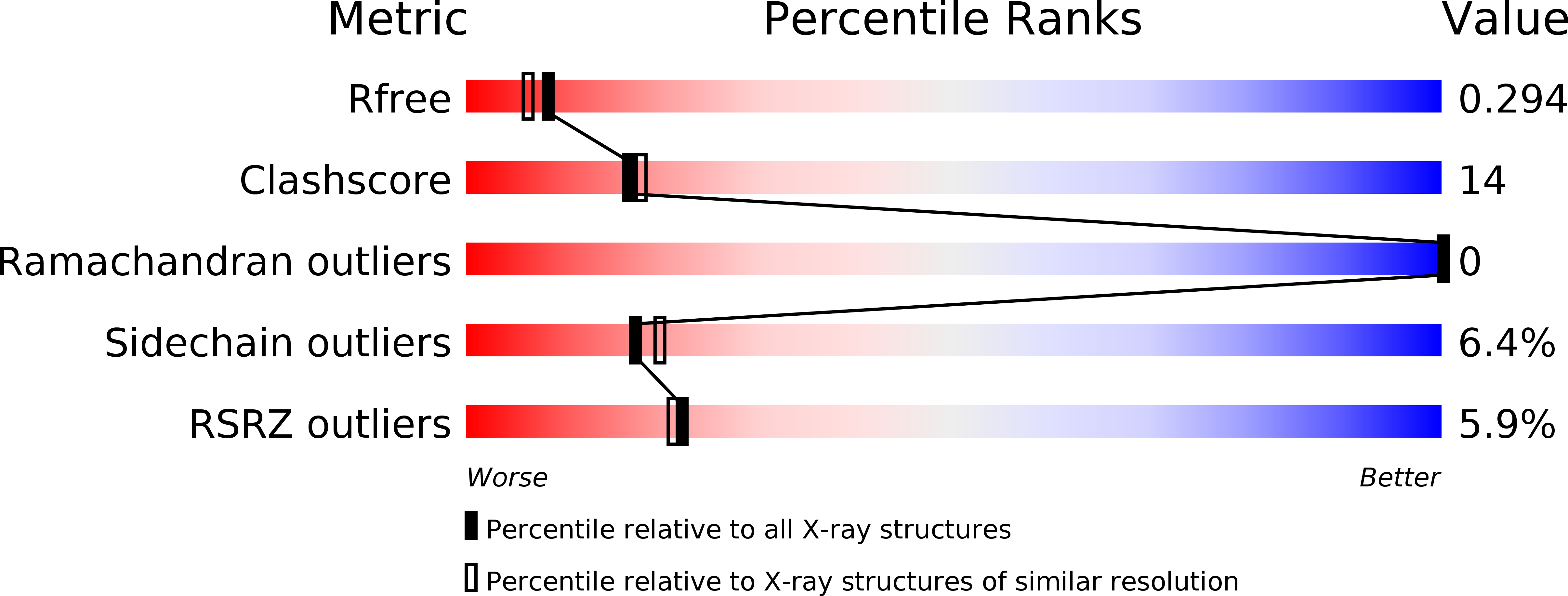
1KWI image
Deposition Date 2002-01-29
Release Date 2002-10-09
Last Version Date 2024-11-20
Entry Detail
PDB ID:
1KWI
Title:
Crystal Structure Analysis of the Cathelicidin Motif of Protegrins
Biological Source:
Source Organism:
Sus scrofa (Taxon ID: 9823)
Method Details:
Experimental Method:
Resolution:
2.19 Å
R-Value Free:
0.29
R-Value Work:
0.22
R-Value Observed:
0.22
Space Group:
P 65 2 2
Macromolecular Entities
Structures with similar UniProt ID
Protein Blast
Polymer Type:polypeptide(L)
Molecule:Protegrin-3 Precursor
Gene (Uniprot):NPG3
Chain IDs:A
Chain Length:101
Number of Molecules:1
Biological Source:Sus scrofa
Primary Citation
Structure of the cathelicidin motif of protegrin-3 precursor: structural insights into the activation mechanism of an antimicrobial protein.
Structure 10 1363 1370 (2002)
PMID: 12377122 DOI: 10.1016/S0969-2126(02)00859-6

Abstact

Cathelicidins are a family of antimicrobial proteins isolated from leucocytes and epithelia cells that contribute to the innate host defense mechanisms in mammalians. Located in the C-terminal part of the holoprotein, the cathelicidin-derived antimicrobial peptide is liberated by a specific protease cleavage. Here, we report the X-ray structure of the cathelicidin motif of protegrin-3 solved by MAD phasing using the selenocysteine-labeled protein. Its overall structure represents a fold homologous to the cystatin family and adopts two native states, a monomer, and a domain-swapped dimer. This crystal structure is the first example of a structural characterization of the highly conserved cathelicidin motif and thus provides insights into the possible mechanism of activation of the antimicrobial protegrin peptide.

Legend

Protein

Chemical

Disease

Primary Citation of related structures