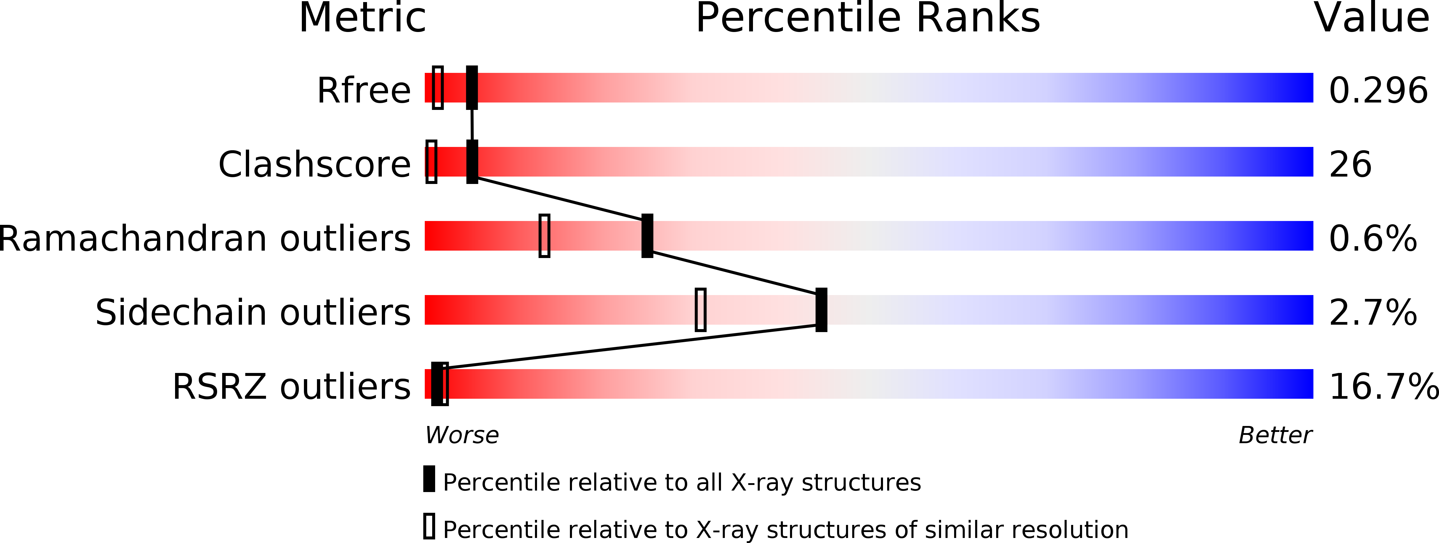
1KWA image
Deposition Date 1998-01-16
Release Date 1998-05-27
Last Version Date 2024-02-14
Entry Detail
PDB ID:
1KWA
Keywords:
Title:
HUMAN CASK/LIN-2 PDZ DOMAIN
Biological Source:
Source Organism:
Homo sapiens (Taxon ID: 9606)
Host Organism:
Method Details:
Experimental Method:
Resolution:
1.93 Å
R-Value Free:
0.29
R-Value Work:
0.25
R-Value Observed:
0.25
Space Group:
C 2 2 21
Macromolecular Entities
Polymer Type:polypeptide(L)
Molecule:HCASK/LIN-2 PROTEIN
Gene (Uniprot):CASK
Chain IDs:A, B
Chain Length:88
Number of Molecules:2
Biological Source:Homo sapiens
Ligand Molecules
Primary Citation
Crystal structure of the hCASK PDZ domain reveals the structural basis of class II PDZ domain target recognition
Nat.Struct.Biol. 5 317 325 (1998)
PMID: 9546224 DOI: 10.1038/nsb0498-317

Abstact

PDZ domain containing proteins assist formation of cell-cell junctions and localization of membrane protein receptors and ion channels. PDZ domains interact with the C-terminal residues of a particular target membrane protein. Based on their binding specificities and sequence homologies, PDZ domains fall into two classes. The first crystal structure of a class II PDZ domain, that of hCASK, has been solved by multi-wavelength anomalous dispersion phasing. Complex formation with the C-terminus of a neighboring non-crystallographically related PDZ domain reveals interactions between hCASK and its ligand. Class II PDZ domains differ from class I domains by formation of a second hydrophobic binding pocket. The C-terminal carboxylate binding loop of the PDZ domain is structurally conserved in both classes suggesting a generalized carboxylate binding motif (h-Gly-h) where h is a hydrophobic residue.

Legend

Protein

Chemical

Disease

Primary Citation of related structures