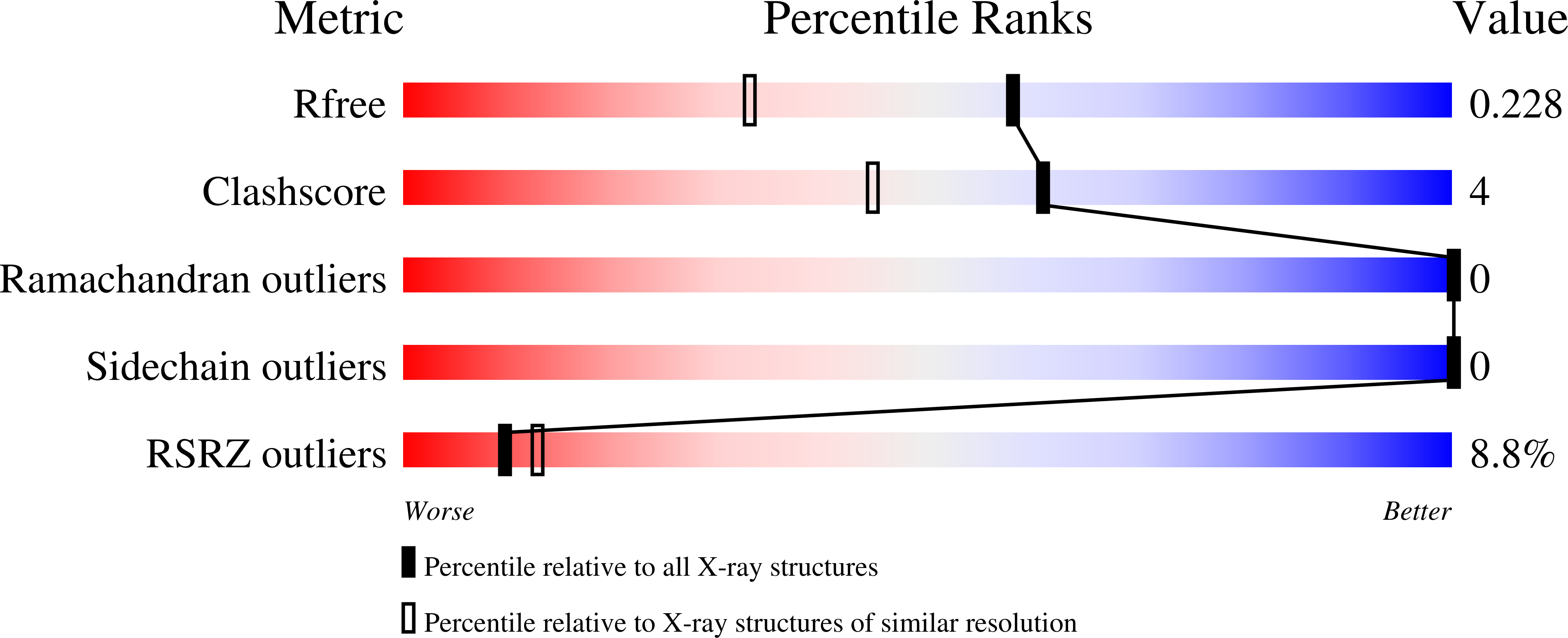
1KW4 image
Deposition Date 2002-01-28
Release Date 2002-06-05
Last Version Date 2024-11-20
Entry Detail
PDB ID:
1KW4
Title:
Polyhomeotic SAM domain structure
Biological Source:
Source Organism:
Host Organism:
Method Details:
Experimental Method:
Resolution:
1.75 Å
R-Value Free:
0.23
R-Value Work:
0.22
R-Value Observed:
0.22
Space Group:
P 65
Macromolecular Entities
Structures with similar UniProt ID
Protein Blast
Polymer Type:polypeptide(L)
Molecule:Polyhomeotic
Gene (Uniprot):ph-p
Mutagens:L51R
Chain IDs:A
Chain Length:89
Number of Molecules:1
Biological Source:Drosophila melanogaster
Modified Residue
Compound ID Chain ID Parent Comp ID Details 2D Image
MSE A MET SELENOMETHIONINE
Primary Citation
The SAM domain of polyhomeotic forms a helical polymer.
Nat.Struct.Biol. 9 453 457 (2002)
PMID: 11992127

Abstact

The polycomb group (PcG) proteins are important in the maintenance of stable repression patterns during development. Several PcG members contain a protein protein interaction module called a SAM domain (also known as SPM, PNT and HLH). Here we report the high-resolution structure of the SAM domain of polyhomeotic (Ph). Ph-SAM forms a helical polymer structure, providing a likely mechanism for the extension of PcG complexes. The structure of the polymer resembles that formed by the SAM domain of another transcriptional repressor, TEL. The formation of these polymer structures by SAM domains in two divergent repressors suggests a conserved mode of repression involving a higher order chromatin structure.

Legend

Protein

Chemical

Disease

Primary Citation of related structures