1KVZ image
Deposition Date 2002-01-28
Release Date 2002-07-28
Last Version Date 2024-10-23
Entry Detail
PDB ID:
1KVZ
Keywords:
Title:
Solution Structure of Cytotoxic RC-RNase4
Biological Source:
Source Organism:
Method Details:
Experimental Method:
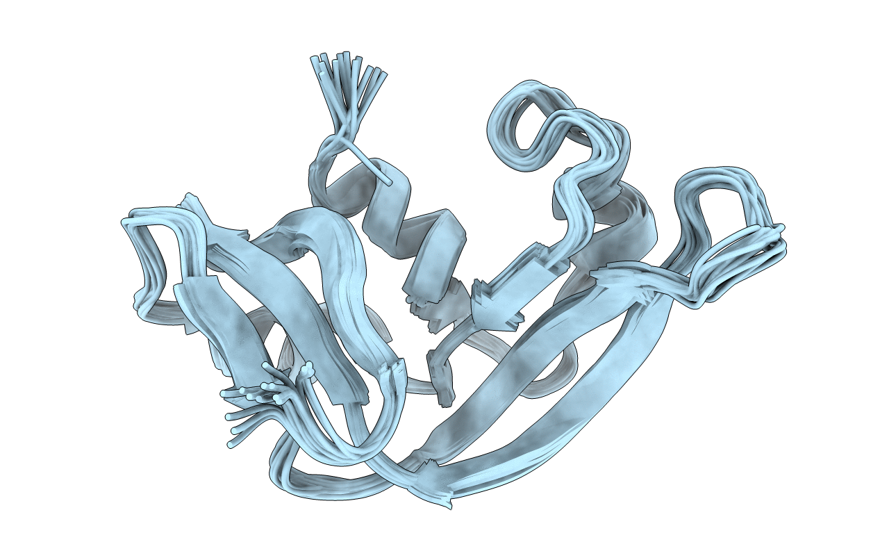
Conformers Calculated:
200
Conformers Submitted:
15
Selection Criteria:
structures with the lowest energy
Macromolecular Entities
Structures with similar UniProt ID
Protein Blast
Polymer Type:polypeptide(L)
Molecule:RC-RNase4
Chain IDs:A
Chain Length:107
Number of Molecules:1
Biological Source:Rana catesbeiana
Ligand Molecules
Primary Citation
Solution Structure of the Cytotoxic RNase 4 from the Oocytes of Bullfrog Rana Catesbeiana
J.MOL.BIOL. 326 1189 1201 (2003)
PMID: 12589762 DOI: 10.1016/S0022-2836(02)01472-9

Abstact

Cytotoxic ribonucleases with antitumor activity are mainly found in the oocytes and early embryos of frogs. Native RC-RNase 4 (RNase 4), consisting of 106 residues linked with four disulfide bridges, is a cytotoxic ribonuclease isolated from oocytes of bullfrog Rana catesbeiana. RNase 4 belongs to the bovine pancreatic ribonuclease (RNase A) superfamily. Recombinant RC-RNase 4 (rRNase 4), which contains an additional Met residue and glutamine instead of pyroglutamate at the N terminus, was found to possess less catalytic and cytotoxic activities than RNase 4. Equilibrium thermal and guanidine-HCl denaturation CD measurements revealed that RNase 4 is more thermally and chemically stable than rRNase 4. However, CD and NMR data showed that there is no gross conformational change between native and recombinant RNase 4. The NMR solution structure of rRNase 4 was determined to comprise three alpha-helices and two sets of antiparallel beta-sheets. Superimposition of each structure with the mean structure yielded an average root mean square deviation (RMSD) of 0.72(+/-0.14)A for the backbone atoms, and 1.42(+/-0.19)A for the heavy atoms in residues 3-105. A comparison of the 3D structure of rRNase 4 with the structurally and functionally related cytotoxic ribonuclease, onconase (ONC), showed that the two H-bonds in the N-terminal pyroglutamate of ONC were not present at the corresponding glutamine residue of rRNase 4. We suggest that the loss of these two H-bonds is one of the key factors responsible for the reductions of the conformational stability, catalytic and cytotoxic activities in rRNase 4. Furthermore, the differences of side-chain conformations of subsite residues among RNase A, ONC and rRNase 4 are related to their distinct catalytic activities and base preferences.

Legend

Protein

Chemical

Disease

Primary Citation of related structures