Deposition Date
2002-01-27
Release Date
2002-03-13
Last Version Date
2024-10-09
Entry Detail
PDB ID:
1KVM
Keywords:
Title:
X-ray Crystal Structure of AmpC WT beta-Lactamase in Complex with Covalently Bound Cephalothin
Biological Source:
Source Organism(s):
Escherichia coli (Taxon ID: 562)
Expression System(s):
Method Details:
Experimental Method:
Resolution:
2.06 Å
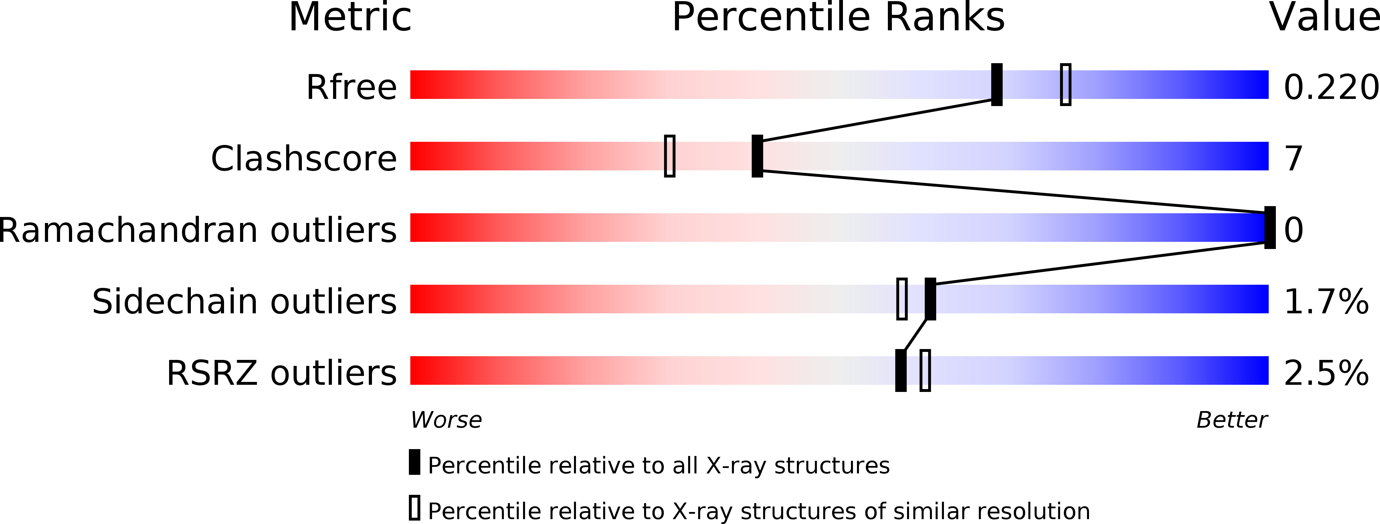
R-Value Free:
0.21
R-Value Work:
0.17
Space Group:
C 1 2 1