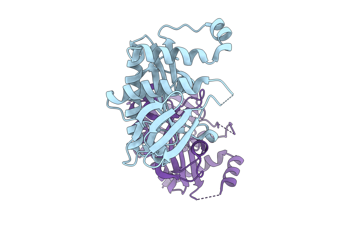
1KUT image
Deposition Date 2002-01-22
Release Date 2002-08-14
Last Version Date 2024-11-20
Entry Detail
PDB ID:
1KUT
Title:
Structural Genomics, Protein TM1243, (SAICAR synthetase)
Biological Source:
Source Organism:
Method Details:
Experimental Method:
Resolution:
2.20 Å
R-Value Free:
0.28
R-Value Work:
0.24
Space Group:
P 1 21 1
Macromolecular Entities
Polymer Type:polypeptide(L)
Molecule:Phosphoribosylaminoimidazole-succinocarboxamide synthase
Gene (Uniprot):purC
Chain IDs:A, B
Chain Length:230
Number of Molecules:2
Biological Source:Thermotoga maritima
Modified Residue
Compound ID Chain ID Parent Comp ID Details 2D Image
MSE A MET SELENOMETHIONINE
Primary Citation
Structure of SAICAR synthase from Thermotoga maritima at 2.2 angstroms reveals an unusual covalent dimer.
Acta Crystallogr.,Sect.F 62 335 339 (2006)
PMID: 16582479 DOI: 10.1107/S1744309106009651

Abstact

As a part of a structural genomics program, the 2.2 angstroms resolution crystal structure of the PurC gene product from Thermotoga maritima has been solved. This 26.2 kDa protein belongs to the phophoribosylaminoimidazole-succinocarboxamide or SAICAR synthase family of enzymes, the members of which are involved in de novo purine biosynthesis. SAICAR synthase can be divided into three subdomains: two alpha+beta regions exhibiting structural homology with ATP-binding proteins and a carboxy-terminal subdomain of two alpha-helices. The asymmetric unit contains two copies of the protein which are covalently linked by a disulfide bond between Cys126(A) and Cys126(B). This 230-amino-acid protein exhibits high structural homology with SAICAR synthase from baker's yeast. The protein structure is described and compared with that of the ATP-SAICAR synthase complex from yeast.

Legend

Protein

Chemical

Disease

Primary Citation of related structures