Deposition Date
2002-01-18
Release Date
2002-02-27
Last Version Date
2024-10-09
Entry Detail
PDB ID:
1KTZ
Keywords:
Title:
Crystal Structure of the Human TGF-beta Type II Receptor Extracellular Domain in Complex with TGF-beta3
Biological Source:
Source Organism(s):
Homo sapiens (Taxon ID: 9606)
Expression System(s):
Method Details:
Experimental Method:
Resolution:
2.15 Å
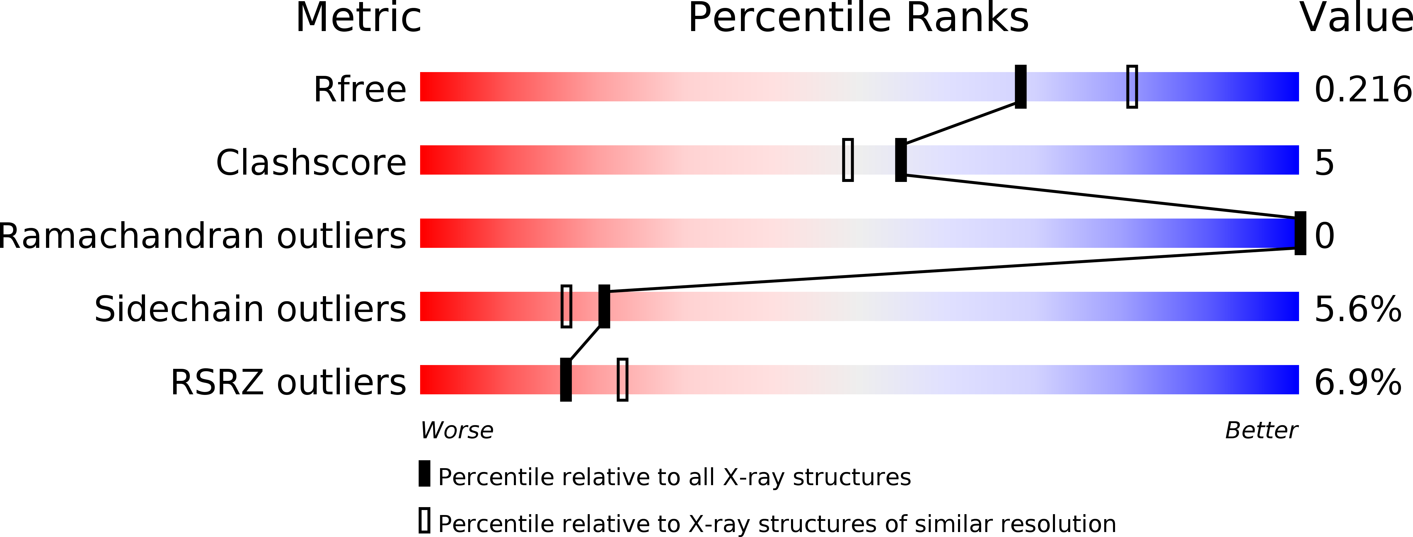
R-Value Free:
0.21
R-Value Work:
0.20
R-Value Observed:
0.20
Space Group:
H 3 2