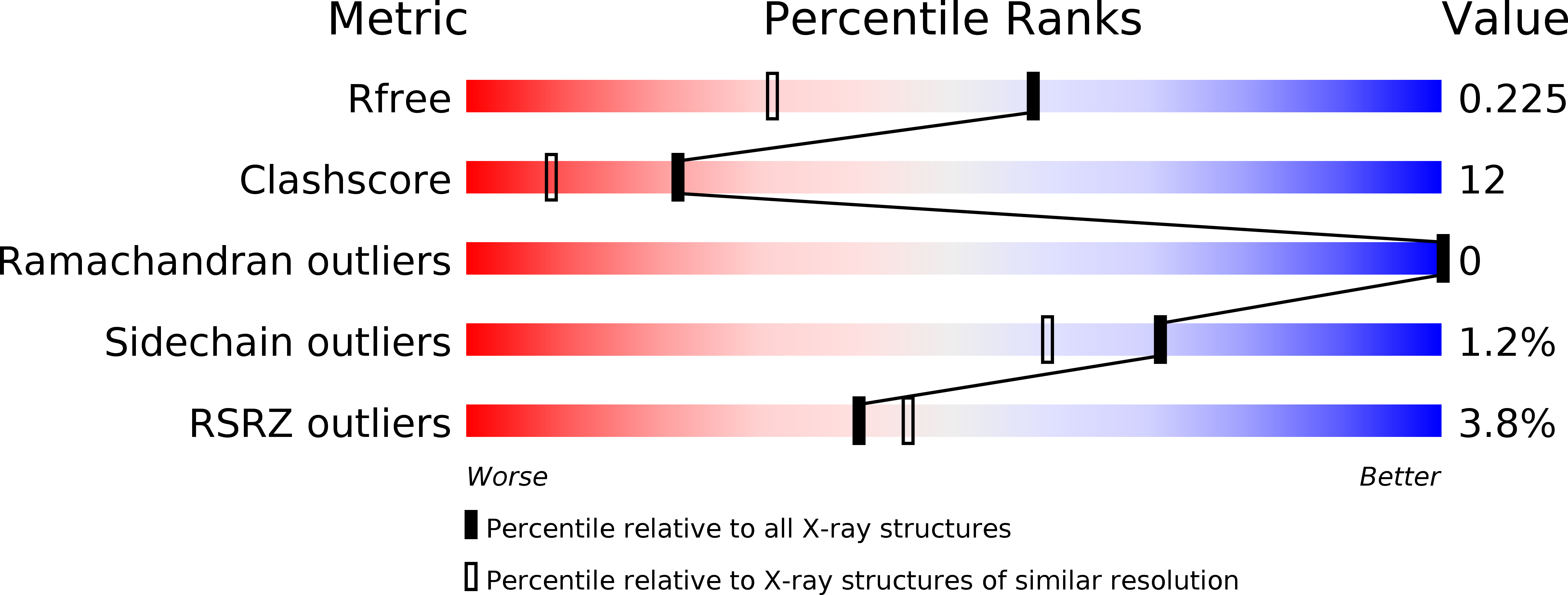
1KSO image
Deposition Date 2002-01-14
Release Date 2002-07-31
Last Version Date 2024-02-14
Entry Detail
PDB ID:
1KSO
Title:
CRYSTAL STRUCTURE OF APO S100A3
Biological Source:
Source Organism:
Homo sapiens (Taxon ID: 9606)
Host Organism:
Method Details:
Experimental Method:
Resolution:
1.70 Å
R-Value Free:
0.22
R-Value Work:
0.19
R-Value Observed:
0.19
Space Group:
P 31
Macromolecular Entities
Structures with similar UniProt ID
Protein Blast
Polymer Type:polypeptide(L)
Molecule:S100 CALCIUM-BINDING PROTEIN A3
Gene (Uniprot):S100A3
Chain IDs:A, B
Chain Length:101
Number of Molecules:2
Biological Source:Homo sapiens
Primary Citation
Metal-free MIRAS phasing: structure of apo-S100A3.
Acta Crystallogr.,Sect.D 58 1255 1261 (2002)
PMID: 12136135 DOI: 10.1107/S0907444902008430

Abstact

S100 proteins are involved in metal-dependent intracellular signalling. Metal-free S100A3, a cysteine-rich Ca(2+)- and Zn(2+)-binding protein, has been crystallized by vapour diffusion under the strict exclusion of oxygen and in the absence of divalent metal ions. Metal binding induces large conformational changes, rendering the apo-S100A3 crystals very sensitive to various metal compounds. Therefore, the structure was solved by MIRAS phasing using potassium iodide and xenon derivatives. Iodide replaces a water molecule at the surface of the S100A3 protein, whereas xenon binds in a hydrophobic cavity at the dimer interface. Despite significant non-isomorphism, the combination of both derivatives was sufficient for structure determination. The overall apo-S100A3 structure resembles the structures of metal-free S100B and S100A6 solution structures. In contrast to the NMR structures, the EF-hand loops are well ordered in the apo-S100A3 crystal structure. In the N-terminal pseudo-EF-hand loop a water molecule occupies the position of the Ca(2+) ion. The C-terminal canonical EF-hand loop shows an extended conformation and a different helix arrangement to other S100/metal complex crystal structures.

Legend

Protein

Chemical

Disease

Primary Citation of related structures