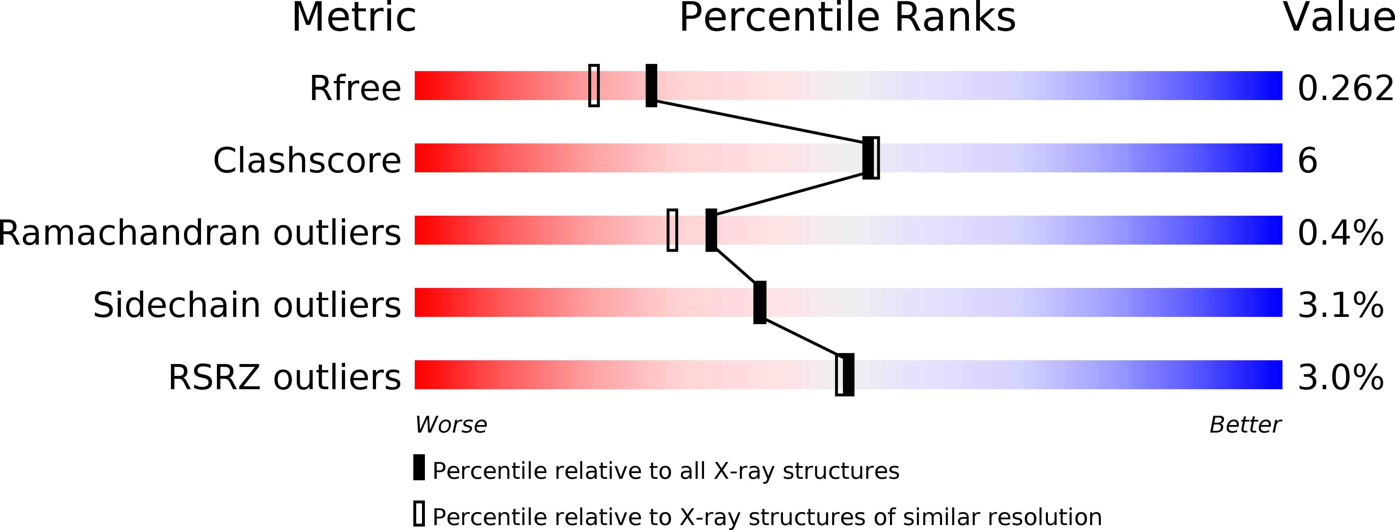
1KSK image
Deposition Date 2002-01-13
Release Date 2002-04-24
Last Version Date 2024-11-06
Entry Detail
PDB ID:
1KSK
Keywords:
Title:
STRUCTURE OF RSUA
Biological Source:
Source Organism:
Escherichia coli (Taxon ID: 562)
Host Organism:
Method Details:
Experimental Method:
Resolution:
2.00 Å
R-Value Free:
0.26
R-Value Work:
0.21
Space Group:
P 21 21 2
Macromolecular Entities
Structures with similar UniProt ID
Protein Blast
Polymer Type:polypeptide(L)
Molecule:RIBOSOMAL SMALL SUBUNIT PSEUDOURIDINE SYNTHASE A
Gene (Uniprot):rsuA
Chain IDs:A
Chain Length:234
Number of Molecules:1
Biological Source:Escherichia coli
Modified Residue
Compound ID Chain ID Parent Comp ID Details 2D Image
MSE A MET SELENOMETHIONINE
Ligand Molecules
Primary Citation
Structure of the 16S rRNA pseudouridine synthase RsuA bound to uracil and UMP.
Nat.Struct.Biol. 9 353 358 (2002)
PMID: 11953756

Abstact

In Escherichia coli, the pseudouridine synthase RsuA catalyzes formation of pseudouridine (psi) at position 516 in 16S rRNA during assembly of the 30S ribosomal subunit. We have determined the crystal structure of RsuA bound to uracil at 2.0 A resolution and to uridine 5'-monophosphate (UMP) at 2.65 A resolution. RsuA consists of an N-terminal domain connected by an extended linker to the central and C-terminal domains. Uracil and UMP bind in a cleft between the central and C-terminal domains near the catalytic residue Asp 102. The N-terminal domain shows structural similarity to the ribosomal protein S4. Despite only 15% amino acid identity, the other two domains are structurally similar to those of the tRNA-specific psi-synthase TruA, including the position of the catalytic Asp. Our results suggest that all four families of pseudouridine synthases share the same fold of their catalytic domain(s) and uracil-binding site.

Legend

Protein

Chemical

Disease

Primary Citation of related structures