1KR7 image
Deposition Date 2002-01-09
Release Date 2002-05-15
Last Version Date 2024-02-14
Entry Detail
PDB ID:
1KR7
Title:
Crystal structure of the nerve tissue mini-hemoglobin from the nemertean worm Cerebratulus lacteus
Biological Source:
Source Organism:
Host Organism:
Method Details:
Experimental Method:
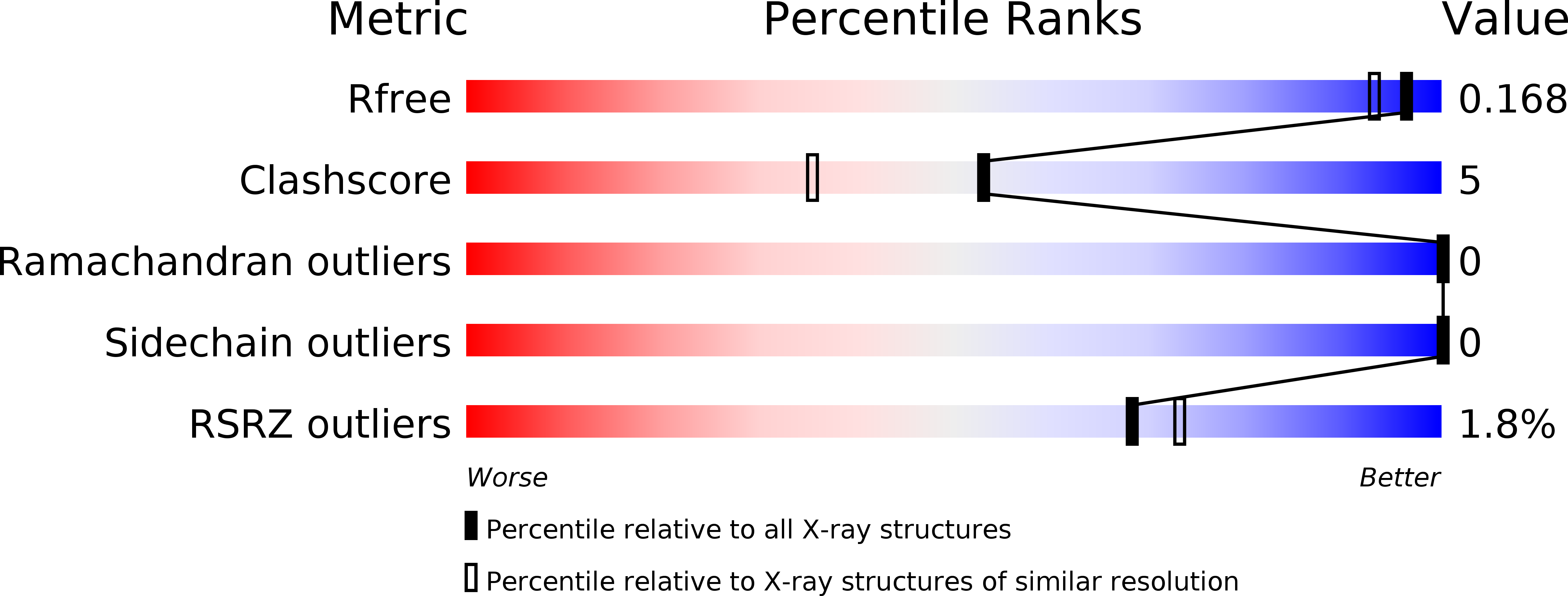
Resolution:
1.50 Å
R-Value Free:
0.18
R-Value Work:
0.15
R-Value Observed:
0.15
Space Group:
P 21 21 21
Macromolecular Entities
Polymer Type:polypeptide(L)
Molecule:Neural globin
Chain IDs:A
Chain Length:110
Number of Molecules:1
Biological Source:Cerebratulus lacteus
Primary Citation
The 109 residue nerve tissue minihemoglobin from Cerebratulus lacteus highlights striking structural plasticity of the alpha-helical globin fold
Structure 10 725 735 (2002)
PMID: 12015154 DOI: 10.1016/S0969-2126(02)00763-3

Abstact

A very short hemoglobin (CerHb; 109 amino acids) binds O(2) cooperatively in the nerve tissue of the nemertean worm Cerebratulus lacteus to sustain neural activity during anoxia. Sequence analysis suggests that CerHb tertiary structure may be unique among the known globin fold evolutionary variants. The X-ray structure of oxygenated CerHb (R factor 15.3%, at 1.5 A resolution) displays deletion of the globin N-terminal A helix, an extended GH region, a very short H helix, and heme solvent shielding based on specific aromatic residues. The heme-bound O(2) is stabilized by hydrogen bonds to the distal TyrB10-GlnE7 pair. Ligand access to heme may take place through a wide protein matrix tunnel connecting the distal site to a surface cleft located between the E and H helices.

Legend

Protein

Chemical

Disease

Primary Citation of related structures