1KQR image
Deposition Date 2002-01-07
Release Date 2002-03-27
Last Version Date 2024-02-14
Entry Detail
PDB ID:
1KQR
Keywords:
Title:
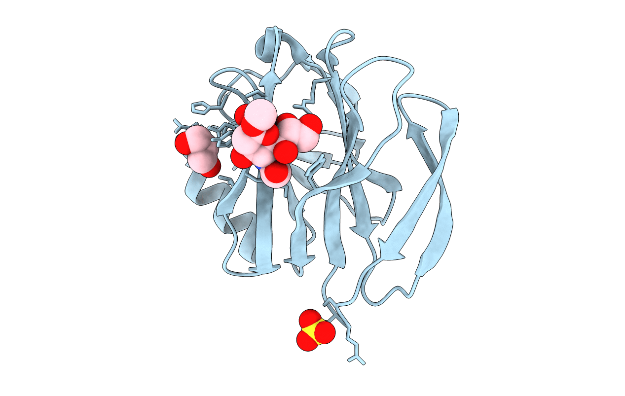
Crystal Structure of the Rhesus Rotavirus VP4 Sialic Acid Binding Domain in Complex with 2-O-methyl-alpha-D-N-acetyl neuraminic acid
Biological Source:
Source Organism:
Method Details:
Experimental Method:
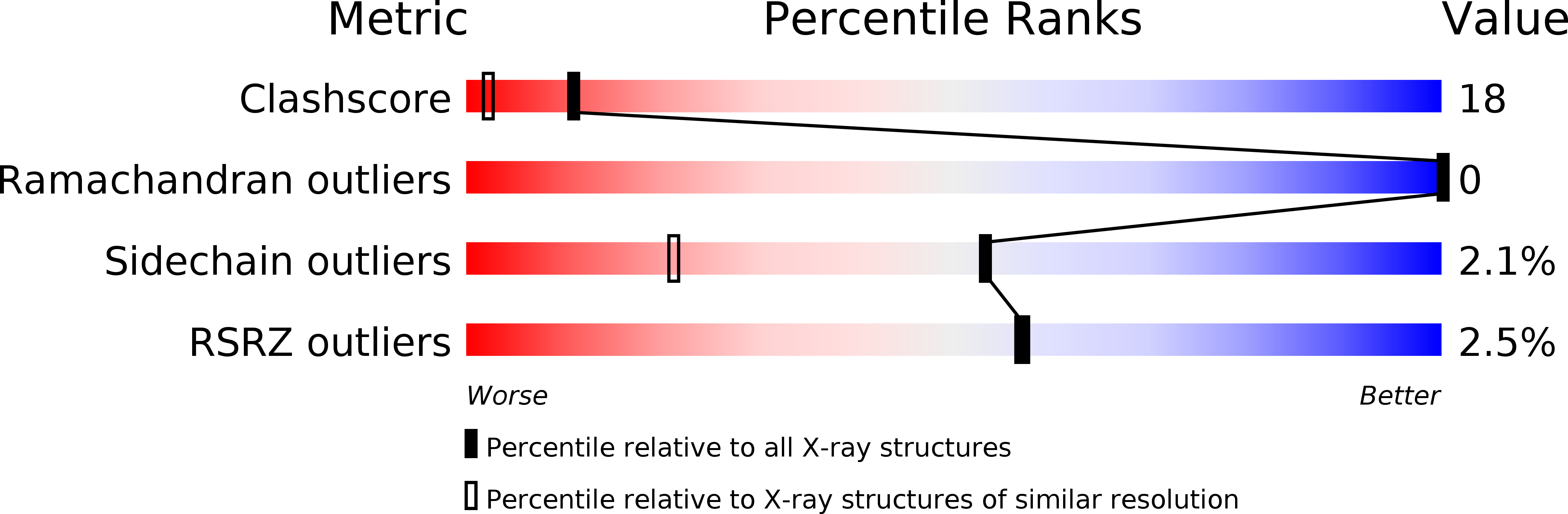
Resolution:
1.40 Å
R-Value Free:
0.18
R-Value Work:
0.16
Space Group:
P 41 21 2
Macromolecular Entities
Polymer Type:polypeptide(L)
Molecule:VP4
Chain IDs:A
Chain Length:179
Number of Molecules:1
Biological Source:Rhesus rotavirus
Primary Citation
The Rhesus Rotavirus VP4 Sialic Acid Binding Domain has a Galectin Fold with a Novel Carbohydrate Binding Site
Embo J. 21 885 897 (2002)
PMID: 11867517 DOI: 10.1093/emboj/21.5.885

Abstact

Cell attachment and membrane penetration are functions of the rotavirus outer capsid spike protein, VP4. An activating tryptic cleavage of VP4 produces the N-terminal fragment, VP8*, which is the viral hemagglutinin and an important target of neutralizing antibodies. We have determined, by X-ray crystallography, the atomic structure of the VP8* core bound to sialic acid and, by NMR spectroscopy, the structure of the unliganded VP8* core. The domain has the beta-sandwich fold of the galectins, a family of sugar binding proteins. The surface corresponding to the galectin carbohydrate binding site is blocked, and rotavirus VP8* instead binds sialic acid in a shallow groove between its two beta-sheets. There appears to be a small induced fit on binding. The residues that contact sialic acid are conserved in sialic acid-dependent rotavirus strains. Neutralization escape mutations are widely distributed over the VP8* surface and cluster in four epitopes. From the fit of the VP8* core into the virion spikes, we propose that VP4 arose from the insertion of a host carbohydrate binding domain into a viral membrane interaction protein.

Legend

Protein

Chemical

Disease

Primary Citation of related structures