1KQD image
Deposition Date 2002-01-04
Release Date 2002-02-13
Last Version Date 2024-02-14
Entry Detail
PDB ID:
1KQD
Keywords:
Title:
Structure of Nitroreductase from E. cloacae Bound with 2e-Reduced Flavin Mononucleotide (FMN)
Biological Source:
Source Organism:
Method Details:
Experimental Method:
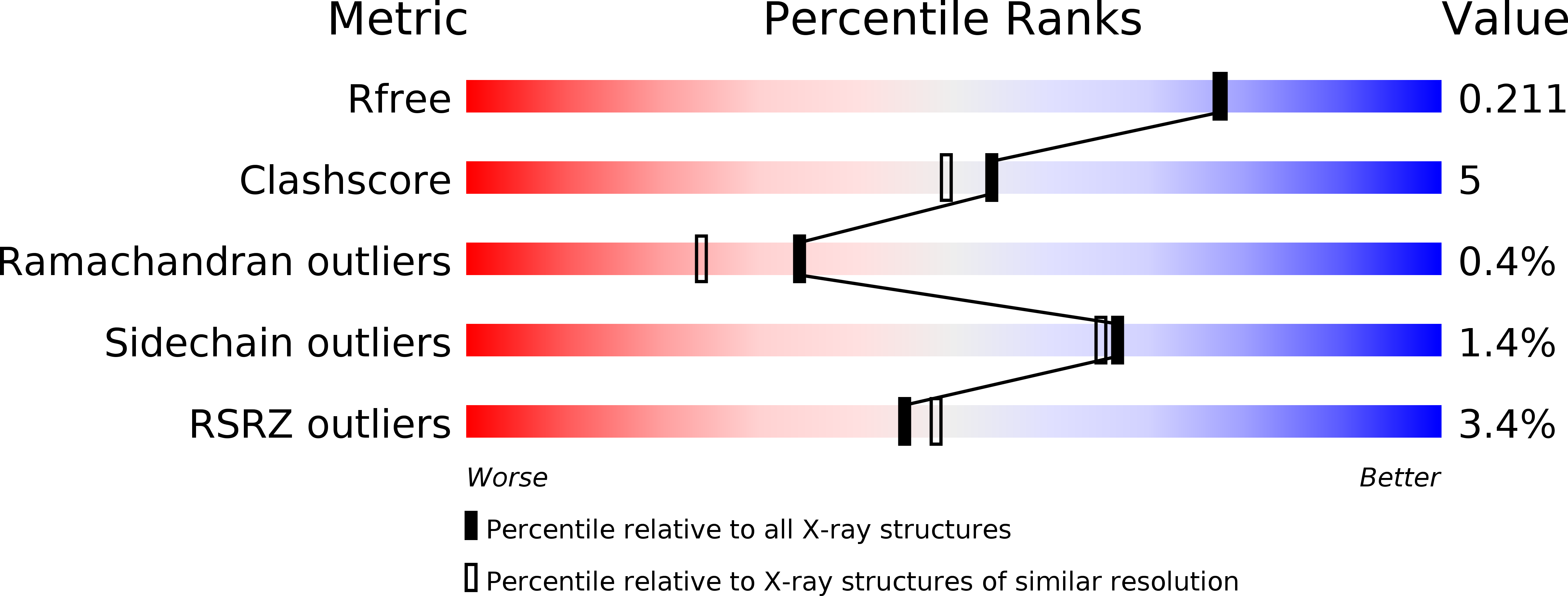
Resolution:
1.90 Å
R-Value Free:
0.21
R-Value Work:
0.18
R-Value Observed:
0.19
Space Group:
P 1 21 1
Macromolecular Entities
Structures with similar UniProt ID
Protein Blast
Polymer Type:polypeptide(L)
Molecule:OXYGEN-INSENSITIVE NAD(P)H NITROREDUCTASE
Gene (Uniprot):nfsB
Chain IDs:A, B, C, D
Chain Length:217
Number of Molecules:4
Biological Source:Enterobacter cloacae
Ligand Molecules
Primary Citation
Structures of nitroreductase in three states: effects of inhibitor binding and reduction.
J.Biol.Chem. 277 11513 11520 (2002)
PMID: 11805110 DOI: 10.1074/jbc.M111334200

Abstact

The crystal structure of the nitroreductase enzyme from Enterobacter cloacae has been determined for the oxidized form in separate complexes with benzoate and acetate inhibitors and for the two-electron reduced form. Nitroreductase is a member of a group of enzymes that reduce a broad range of nitroaromatic compounds and has potential uses in chemotherapy and bioremediation. The monomers of the nitroreductase dimer adopt an alpha+beta fold and together bind two flavin mononucleotide prosthetic groups at the dimer interface. In the oxidized enzyme, the flavin ring system adopts a strongly bent (16 degrees ) conformation, and the bend increases (25 degrees ) in the reduced form of the enzyme, roughly the conformation predicted for reduced flavin free in solution. Because free oxidized flavin is planar, the induced bend in the oxidized enzyme may favor reduction, and it may also account for the characteristic inability of the enzyme to stabilize the one electron-reduced semiquinone flavin, which is also planar. Both inhibitors bind over the pyrimidine and central rings of the flavin in partially overlapping sites. Comparison of the two inhibitor complexes shows that a portion of helix H6 can flex to accommodate the differently sized inhibitors suggesting a mechanism for accommodating varied substrates.

Legend

Protein

Chemical

Disease

Primary Citation of related structures